Pertussis (Bordetella pertussis and Bordetella parapertussis)

Published on 22/03/2015 by admin

Filed under Pediatrics

Last modified 22/04/2025

Print this page

rate 1 star rate 2 star rate 3 star rate 4 star rate 5 star
Your rating: none, Average: 2 (1 votes)

This article have been viewed 1830 times

Chapter 189 Pertussis (Bordetella pertussis and Bordetella parapertussis)

Pertussis is an acute respiratory tract infection that was well described initially in the 1500s. Sydenham first used the term pertussis, meaning intense cough, in 1670; it is preferable to whooping cough because most infected individuals do not “whoop.”

Epidemiology

There are 60 million cases of pertussis each year worldwide, resulting in >500,000 deaths. Before vaccination was available, pertussis was the leading cause of death due to communicable disease among children <14 yr of age in the USA, with 10,000 deaths annually. Widespread use of pertussis vaccine led to a >99% decline in cases. The pivotal role of vaccination in disease control is reflected in the continued high incidence of pertussis in developing countries and the resurgence in other countries where vaccine coverage is low or where less potent vaccine may have been used.

After the low number of 1,010 cases in the USA reported in 1976, there was an increase in annual pertussis incidence to 1.2 cases/100,000 population from 1980 through 1989, with epidemic pertussis in many states in 1989-1990, 1993, and 1996. Since then pertussis has become increasingly endemic, with less cycling or seasonality and with shifting burden of disease to young infants, adolescents, and adults. By 2004, the incidence of reported pertussis in the USA was 8.9 cases/100,000 population, and the number of cases (25,827) was the highest reported since 1959. Of these cases, 10% occurred among infants <6 mo of age (incidence of 136.5/100,000 population). A total of 40 pertussis-related deaths were reported in 2005, and 16 in 2006; >90% occurred among young infants. Approximately 60% of cases currently are in adolescents and adults. Pertussis is the only vaccine-preventable disease for which universal immunization in the USA is recommended that continues to be endemic. Prospective and serologic studies suggest that pertussis is underrecognized, especially among adolescents and adults, in whom the actual number of cases is estimated to be 600,000 annually. A number of studies have documented pertussis in 13-32% of adolescents and adults with cough illness for >7 days.

Pertussis is extremely contagious, with attack rates as high as 100% in susceptible individuals exposed to aerosol droplets at close range. B. pertussis does not survive for prolonged periods in the environment. Chronic carriage by humans is not documented. After intense exposure as in households, the rate of subclinical infection is as high as 80% in fully immunized or previously infected individuals. When carefully sought, a symptomatic source case can be found for most patients.

Neither natural disease nor vaccination provides complete or lifelong immunity against pertussis re-infection or disease. Protection against typical disease begins to wane 3-5 yr after vaccination and is unmeasurable after 12 yr. Subclinical re-infection undoubtedly has contributed significantly to immunity against disease ascribed previously to both vaccine and prior infection. Despite history of disease or complete immunization, outbreaks of pertussis have occurred in the elderly, in nursing homes, in residential facilities with limited exposures, in highly immunized suburbia, and in preadolescents, adolescents, and adults with significant time since immunization. Possible explanations for change in epidemiology include waning immunity post immunization, an aging cohort who received less effective vaccine, and increased awareness and diagnosis. Without natural re-infection with B. pertussis or repeated booster vaccinations, adolescents and adults are susceptible to clinical disease if exposed, and mothers provide little if any passive protection to young infants. Coughing adolescents and adults (usually not recognized as having pertussis) are the major reservoir for B. pertussis and are the usual sources of infection for infants and children.

Clinical Manifestations

Classically, pertussis is a prolonged disease, divided into catarrhal, paroxysmal, and convalescent stages. The catarrhal stage (1-2 wk) begins insidiously after an incubation period ranging from 3-12 days with nondistinctive symptoms of congestion and rhinorrhea variably accompanied by low-grade fever, sneezing, lacrimation, and conjunctival suffusion. As initial symptoms wane, coughing marks the onset of the paroxysmal stage (2-6 wk). The cough begins as a dry, intermittent, irritative hack and evolves into the inexorable paroxysms that are the hallmark of pertussis. A well-appearing, playful toddler with insignificant provocation suddenly expresses an anxious aura and may clutch a parent or comforting adult before beginning a machine-gun burst of uninterrupted cough on a single exhalation, chin and chest held forward, tongue protruding maximally, eyes bulging and watering, face purple, until coughing ceases and a loud whoop follows as inspired air traverses the still partially closed airway. Post-tussive emesis is common, and exhaustion is universal. The number and severity of paroxysms escalate over days to a week and remain at that plateau for days to weeks. At the peak of the paroxysmal stage, patients may have more than 1 episode hourly. As the paroxysmal stage fades into the convalescent stage (≥2 wk), the number, severity, and duration of episodes diminish.

Infants <3 mo of age do not display the classic stages. The catarrhal phase lasts only a few days or is unnoticed, and then, after the most insignificant startle from a draft, light, sound, sucking, or stretching, a well-appearing young infant begins to choke, gasp, gag, and flail the extremities, with face reddened. Cough may not be prominent, especially in the early phase. Whoop infrequently occurs in infants <3 mo of age who at the end of a paroxysm lack stature or muscular strength to create sudden negative intrathoracic pressure. Apnea and cyanosis can follow a coughing paroxysm, or apnea can occur without a cough. Apnea may be the only symptom. Apnea and cyanosis both are more common with pertussis than with neonatal viral infections, including respiratory syncytial virus (RSV). The paroxysmal and convalescent stages in young infants are lengthy. Paradoxically, in infants, cough and whooping may become louder and more classic in convalescence. Convalescence includes intermittent paroxysmal coughing throughout the 1st yr of life, including “exacerbations” with subsequent respiratory illnesses; these are not due to recurrent infection or reactivation of B. pertussis.

Adolescents and previously immunized children have foreshortening of all stages of pertussis. Adults have no distinct stages. Classically, adolescents and adults describe a sudden feeling of strangulation followed by uninterrupted coughs, feeling of suffocation, bursting headache, diminished awareness, and then a gasping breath, usually without a whoop. Post-tussive emesis and intermittency of paroxysms separated by hours of well-being are specific clues to the diagnosis in adolescents and adults. At least 30% of older individuals with pertussis have nonspecific cough illness, distinguished only by duration, which is usually >21 days.

Findings on physical examination generally are uninformative. Signs of lower respiratory tract disease are not expected unless complicating secondary bacterial pneumonia is present. Conjunctival hemorrhages and petechiae on the upper body are common.

Diagnosis

Pertussis should be suspected in any individual who has pure or predominant complaint of cough, especially if the following features are absent: fever, malaise or myalgia, exanthem or enanthem, sore throat, hoarseness, tachypnea, wheezes, and rales. For sporadic cases, a clinical case definition of cough of ≥14 days’ duration with at least 1 associated symptom of paroxysms, whoop, or post-tussive vomiting has a sensitivity of 81% and a specificity of 58% for culture confirmation. Pertussis should be suspected in older children whose cough illness is escalating at 7-10 days and whose coughing episodes are not continuous. Pertussis should be suspected in infants <3 mo of age with gagging, gasping, apnea, cyanosis, or an apparent life-threatening event (ALTE). Sudden infant death is occasionally caused by B. pertussis.

Adenoviral infections are usually distinguishable by associated features, such as fever, sore throat, and conjunctivitis. Mycoplasma causes protracted episodic coughing, but patients usually have a history of fever, headache, and systemic symptoms at the onset of disease as well as more continuous cough and frequent finding of rales on auscultation of the chest. Epidemics of Mycoplasma and B. pertussis in young adults can be difficult to distinguish on clinical grounds. Although pertussis is often included in the laboratory evaluation of young infants with afebrile pneumonia, B. pertussis is not associated with staccato cough (breath with every cough), purulent conjunctivitis, tachypnea, rales or wheezes that typify infection due to Chlamydia trachomatis, or predominant lower respiratory tract signs that typify infection due to RSV. Unless an infant with pertussis has secondary pneumonia (and then appears ill), the findings on examination between paroxysms are entirely normal, including respiratory rate.

Leukocytosis (15,000-100,000 cells/mm3) due to absolute lymphocytosis is characteristic in the catarrhal stage. Lymphocytes are of T- and B-cell origin and are normal small cells, rather than the large atypical lymphocytes seen with viral infections. Adults, partially immune children, and, occasionally, young infants have less impressive lymphocytosis. Absolute increase in neutrophils suggests a different diagnosis or secondary bacterial infection. Eosinophilia is not a manifestation of pertussis. A severe course and death are correlated with extreme leukocytosis (median peak white blood cell count in fatal vs nonfatal cases, 94 vs 18 × 109 cells/L, respectively) and thrombocytosis (median peak platelet count in fatal vs nonfatal cases, 782 vs 556 × 109/L, respectively). Mild hyperinsulinemia and reduced glycemic response to epinephrine have been demonstrated, although hypoglycemia is reported only occasionally. Chest radiographic findings are only mildly abnormal in the majority of hospitalized infants, showing perihilar infiltrate or edema (sometimes with a butterfly appearance) and variable atelectasis. Parenchymal consolidation suggests secondary bacterial infection. Pneumothorax, pneumomediastinum, and subcutaneous emphysema can be seen occasionally.

All current methods for confirmation of infection due to B. pertussis have limitations in sensitivity, specificity, or practicality. Isolation of B. pertussis in culture remains the gold standard for diagnosis. Careful attention must be directed to specimen collection, transport, and isolation technique. The specimen is obtained with deep nasopharyngeal aspiration or with the use of a flexible swab, preferably a Dacron or calcium alginate–tipped swab, held in the posterior nasopharynx for 15-30 sec (or until cough occurs). A 1.0% casamino acid liquid is acceptable for holding a specimen up to 2 hr; Stainer-Scholte broth or Regan-Lowe semisolid transport medium is used for longer transport periods, up to 4 days. The preferred isolation media are Regan-Lowe charcoal agar with 10% horse blood and 5-40 µg/mL cephalexin and Stainer-Scholte media with cyclodextrin resins. Cultures are incubated at 35-37°F in a humid environment and examined daily for 7 days for slow-growing, tiny, glistening colonies. Direct fluorescent antibody (DFA) testing of potential isolates using specific antibody for B. pertussis and B. parapertussis maximizes recovery rates. Direct testing of nasopharyngeal secretions by DFA is a rapid test but is reliable only in laboratories with continuous experience. Polymerase chain reaction (PCR) analysis to test nasopharyngeal wash specimens has a sensitivity similar to that of culture and averts difficulties of isolation, but a standardized validated test is not yet available universally. Results of DFA, culture, and PCR are all expected to be positive in unimmunized, untreated children during the catarrhal and early paroxysmal stages of disease. Less than 10% of any of these test results are positive in partially or remotely immunized individuals tested in the paroxysmal stage. Serologic tests for detection of antibodies to B. pertussis antigens in acute and convalescent samples are the most sensitive tests in immunized individuals and are useful epidemiologically. A single serum sample showing immunoglobulin G (IgG) antibody to pertussis toxin elevated >2 standard deviations above the mean of the immunized population (≈100 EU/mL) indicates recent infection. Standardization of tests and cut point for a positive result are currently being investigated. Tests for IgA and IgM pertussis antibody, or antibody to antigens other than PT, are not reliable methods for diagnosis of pertussis.

Treatment

Goals of therapy are to limit the number of paroxysms, to observe the severity of the cough, to provide assistance when necessary, and to maximize nutrition, rest, and recovery without sequelae (Table 189-1). Infants <3 mo of age with suspected pertussis are always admitted to hospital, as are those between 3 and 6 mo of age unless witnessed paroxysms are not severe, and patients of any age if significant complications occur. Prematurely born young infants and children with underlying cardiac, pulmonary, muscular, or neurologic disorders have a high risk for severe, potentially fatal disease.

The specific, limited goals of hospitalization are to: (1) assess progression of disease and likelihood of life-threatening events at peak of disease; (2) prevent or treat complications; and (3) educate parents in the natural history of the disease and in care that will be given at home. Heart rate, respiratory rate, and pulse oximetry are monitored continuously with alarm settings so that paroxysms can be witnessed and recorded by health care personnel. Detailed cough records and documentation of feeding, vomiting, and weight change provide data to assess severity. Typical paroxysms that are not life threatening have the following features: duration <45 sec; red but not blue color change; tachycardia, bradycardia (not <60 beats/min in infants), or oxygen desaturation that spontaneously resolves at the end of the paroxysm; whooping or strength for self-rescue at the end of the paroxysm; self-expectorated mucus plug; and post-tussive exhaustion but not unresponsiveness. Assessing the need to provide oxygen, stimulation, or suctioning requires skilled personnel who can document an infant’s ability for self-rescue but who will intervene rapidly and expertly when necessary. The benefit of a quiet, dimly lighted, undisturbed, comforting environment cannot be overestimated or forfeited in a desire to monitor and intervene. Feeding children with pertussis is challenging. The risk of precipitating cough by nipple feeding does not warrant nasogastric, nasojejunal, or parenteral alimentation in most infants. The composition or thickness of formula does not affect the quality of secretions, cough, or retention. Large-volume feedings are avoided.

Within 48-72 hr, the direction and severity of disease are obvious, usually from analysis of recorded information. Many infants have marked improvement upon hospitalization and antibiotic therapy, especially if they are hospitalized early in the course of disease or have been removed from aggravating environmental smoke, excessive stimulation, or a dry or polluting heat source. Hospital discharge is appropriate if over a 48-hr period disease severity is unchanged or diminished, no intervention is required during paroxysms, nutrition is adequate, no complication has occurred, and parents are adequately prepared for care at home. Apnea and seizures occur in the incremental phase of illness and in patients with complicated disease. Portable oxygen, monitoring, or suction apparatus should not be needed at home.

Infants who have apnea, paroxysms that repeatedly lead to life-threatening events despite passive delivery of oxygen, or respiratory failure require intubation, pharmaceutically induced paralysis, and ventilation.

Antibiotics

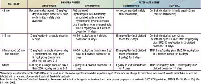
An antimicrobial agent is always given when pertussis is suspected or confirmed, primarily to limit the spread of infection and secondarily for possible clinical benefit. Macrolides are preferred agents and are similar to one another in terms of in vitro activity (Table 189-2). Resistance has been reported rarely. A 7- to 10-fold relative risk for infantile hypertrophic pyloric stenosis (IHPS) has been reported in neonates treated with orally administered erythromycin. Azithromycin is the preferred agent for most patients and particularly in neonates, although cases of IHPS have followed its use. All infants <1 mo of age treated with any macrolide should be monitored for symptoms of pyloric stenosis. Benefits of post-exposure prophylaxis for infants far outweigh risk of IHPS.

Care of Household and Other Close Contacts

A macrolide agent should be given promptly to all household contacts and other close contacts, such as those in daycare, regardless of age, history of immunization, and symptoms (see Table 189-2). The same age-related drugs and doses used for prophylaxis are used for treatment. Visitation and movement of coughing family members in the hospital must be assiduously controlled until erythromycin has been taken for 5 days. In close contacts <7 yr of age who have received fewer than 4 doses of pertussis-containing vaccines, vaccination should be initiated or continued to complete the recommended series. Children <7 yr of age who received a 3rd dose >6 mo before exposure or a 4th dose ≥3 yr before exposure should receive a booster dose. Individuals ≥9 yr of age should be given a Tdap (adolescent/adult formulation of tetanus and diphtheria toxoids and acellular pertussis) booster if they have not previously received Tdap. Unmasked health care personnel (HCP) exposed to untreated cases should be evaluated for post-exposure prophylaxis and follow-up. Coughing HCP with or without known exposure to pertussis should be promptly evaluated for pertussis.

Complications

Infants <6 mo of age have excessive mortality and morbidity; infants <2 mo of age have the highest reported rates of pertussis-associated hospitalization (82%), pneumonia (25%), seizures (4%), encephalopathy (1%), and death (1%). Infants <4 mo of age account for 90% of cases of fatal pertussis. Preterm birth and young maternal age are significantly associated with fatal pertussis. Neonates with pertussis have substantially longer hospitalizations, greater need for oxygen, and greater need for mechanical ventilation than neonates with viral respiratory infection.

The principal complications of pertussis are apnea, secondary infections (such as otitis media and pneumonia), and physical sequelae of forceful coughing. Fever, tachypnea or respiratory distress between paroxysms, and absolute neutrophilia are clues to pneumonia. Expected pathogens include Staphylococcus aureus, Streptococcus pneumoniae, and bacteria of oropharyngeal flora. Increased intrathoracic and intra-abdominal pressure during coughing can result in conjunctival and scleral hemorrhages, petechiae on the upper body, epistaxis, hemorrhage in the central nervous system (CNS) and retina, pneumothorax and subcutaneous emphysema, and umbilical and inguinal hernias. Laceration of the lingual frenulum occurs occasionally.

The need for intensive care and mechanical ventilation is usually limited to infants <3 mo of age and infants with underlying conditions. Respiratory failure due to apnea may precipitate need for intubation and ventilation through the days when disease peaks; prognosis is good. Progressive pulmonary hypertension in very young infants and secondary bacterial pneumonia are severe complications of pertussis and are the usual causes of death. Pulmonary hypertension and cardiogenic shock with fatal outcome are associated with extreme elevations of lymphocyte and platelet counts. Autopsies in fatal cases show luminal aggregates of leukocytes in the pulmonary vasculature. Extracorporeal membrane oxygenation of infants with pertussis in whom mechanical ventilation failed has been associated with >80% fatality (questioning the advisability of this procedure). Exchange transfusion or leukopheresis, however, has been associated with drop in lymphocyte and platelet counts, with recovery in several reported cases.

CNS abnormalities occur at a relatively high frequency in pertussis and are almost always a result of hypoxemia or hemorrhage associated with coughing or apnea in young infants. Apnea or bradycardia or both may result from apparent laryngospasm or vagal stimulation just before a coughing episode, from obstruction during an episode, or from hypoxemia following an episode. Lack of associated respiratory signs in some young infants with apnea raises the possibility of a primary effect of PT on the CNS. Seizures are usually a result of hypoxemia, but hyponatremia from excessive secretion of antidiuretic hormone during pneumonia can occur. The only neuropathology documented in humans is parenchymal hemorrhage and ischemic necrosis.

Bronchiectasis has been reported rarely after pertussis. Children who have pertussis before the age of 2 yr may have abnormal pulmonary function into adulthood.

Prevention

Universal immunization of children with pertussis vaccine, beginning in infancy with periodic reinforcing doses through adolescence and adulthood, is central to the control of pertussis (Chapter 165). There is no serologic correlate of protection from B. pertussis.

DTaP Vaccines

Several diphtheria and tetanus toxoids combined with acellular pertussis (DTaP) vaccines or combination products currently are licensed in the USA for children <7 yr of age. DTaP vaccines have fewer adverse effects than the vaccines containing whole-cell pertussis (DTP), which continue to be given to infants and children in many other countries. Acellular pertussis vaccines all contain inactivated PT and 2 or more other bacterial components (FHA, Pn, and Fim 2 and 3). Clinical efficacy against severe pertussis, defined as paroxysmal cough >21 days, is 80-85%. Mild local and systemic adverse events as well as more serious events (including high fever, persistent crying of ≥3 hr in duration, hypotonic hyporesponsive episodes, and seizures) occur significantly less frequently among infants who receive DTaP than in those who receive DTP vaccine. DTaP-containing vaccines can be administered simultaneously with any other vaccines used in standard schedules for children.

Four doses of DTaP should be administered during the 1st 2 years of life, generally at ages 2, 4, 6, and 15-18 mo of age. The 4th dose may be administered as early as 12 mo of age, provided that 6 mo have elapsed since the 3rd dose. The 5th dose of DTaP is recommended for children at 4-6 yr of age; a 5th dose is not necessary if the 4th dose in the series is administered on or after the 4th birthday. A birth dose of DTaP is not effective, but commencement of vaccination at 6 wk of age, with monthly doses through the 3rd dose, can be considered in high-risk settings.

When feasible, the same DTaP product is recommended for all doses of the primary vaccination series. Local reactions increase in rate and severity with successive doses of DTaP, although never reaching the magnitude of reactions following similar doses of DTP. Swelling of the entire thigh or upper arm has been reported in 2-3% of vaccinees after the 4th or 5th doses of a variety of DTaP products. Swelling may be accompanied by pain, erythema, and fever. Limitation of activity is less than might be expected. Swelling subsides spontaneously without sequelae. The pathogenesis is unknown. Extensive limb swelling after the 4th dose of DTaP usually is not associated with a similar reaction to the 5th dose and is not a contraindication to subsequent dose(s).

Exempting children from pertussis immunization should be considered only in the narrow limits as recommended. Exemptors have been shown to have significantly increased risk for pertussis and to play a role in outbreaks of pertussis among immunized populations. Although well-documented pertussis confers short-term protection, the duration of protection is unknown; immunization should be completed on schedule in children diagnosed with pertussis. Improper vaccine storage reduces immunity.

Tdap Vaccines

Two tetanus toxoid, reduced diphtheria toxoid, and acellular pertussis vaccine, adsorbed (Tdap) products were licensed in 2005 and were recommended universally in 2006 for use in individuals 11-18 years of age and in older individuals as a single-dose booster vaccine to provide protection against tetanus, diphtheria, and pertussis. Because of heightened risk in adolescents for pertussis and the evidence of association of pertussis with young infants of adolescent mothers, the American Academy of Pediatrics (AAP) includes pregnant adolescents who are in their second or third trimester in Tdap recommendations. The preferred age for Tdap vaccination is 11-12 yr. All adolescents 11-18 yr of age who received Td, but not Tdap, should receive a single dose of Tdap to provide protection against pertussis. There is no minimum interval required between vaccines containing tetanus or diphtheria toxoid and Tdap. There is no contraindication to concurrent administration of any other indicated vaccine. In 2010, Tdap was recommended for children 7-10 yr old with incomplete pertussis vaccination prior to age 7 yr as well as for those ≥65 yr who have contact with infants. An important objective of administering Tdap is to protect adolescents and adults against pertussis in order to control endemic and epidemic spread to young infants who have not completed primary immunization and are at high risk for pertussis and its complications. In provinces and territories in Canada where Tdap was administered to 14 to 16 yr old adolescents, marked reduction in pertussis has been documented in adolescents and younger age groups, possibly as a result of herd protection. In 2008 and pending further data, the CDC recommendations for pregnant adult women showed preference for the cocoon strategy (immediate postpartum Tdap immunization of mother and all household and other contacts of the infant) over maternal immunization during pregnancy.

Bibliography

American Academy of Pediatrics, Committee on Infectious Diseases. Prevention of pertussis among adolescents: recommendations for use of tetanus toxoid, reduced diphtheria toxoid, and acellular pertussis (Tdap) vaccine. Pediatrics. 2006;117:965-978.

Castagnini LA, Munoz FM. Clinical characteristics and outcomes of neonatal pertussis: a comparative study. J Pediatr. 2010;156:498-500.

Centers for Disease Control and Prevention. Local health department costs associated with response to a school-based pertussis outbreak—Omaha, Nebraska, Sept-Nov, 2008. MMWR. 2011;60(1):5-8.

Centers for Disease Control and Prevention. Use of mass Tdap vaccination to control an outbreak of pertussis in a high school—Cook County, Illinois, September 2006—January 2007. MMWR Morbid Mortal Wkly Rep. 2008;57:796-799.

Centers for Disease Control and Prevention. Recommended antimicrobial agents for treatment and postexposure prophylaxis of pertussis: 2005 CDC guidelines. MMWR Morbid Mortal Wkly Rep. 2005;54:1-16.

Centers for Disease Control and Prevention. Preventing tetanus, diphtheria, and pertussis among adolescents: use of tetanus toxoid, reduced diphtheria toxoid and acellular pertussis vaccines. Recommendations of the Advisory Committee on Immunization Practices (ACIP). MMWR Morbid Mortal Wkly Rep. 2006;55(RR-3):1-34.

Centers for Disease Control and Prevention. Preventing tetanus, diphtheria, and pertussis among pregnant and postpartum women and their infants: recommendations of the Advisory Committee on Immunization Practices (ACIP). MMWR Morbid Mortal Wkly Rep. 2008;57:1-51.

Cornia PB, Hersh AL, Lipsky BA, et al. Does this coughing adolescent or adult patient have pertussis? JAMA. 2010;304(8):890-896.

Dylag AM, Shah SI. Administration of tetanus, diphtheria, and acellular pertussis vaccine to parents of high-risk infants in the neonatal intensive care unit. Pediatrics. 2008;122:e550-e555.

Glanz JM, McClure DL, Magid DJ, et al. Parental refusal of pertussis vaccination is associated with an increased risk of pertussis infection in children. Pediatrics. 2009;123:1446-1451.

Greenberg DP, Doemland M, Bettinger JA, et al. Epidemiology of pertussis and Haemophilus influenzae type b disease in Canada with exclusive use of a diphtheria-tetanus-acellular pertussis-inactivated poliovirus—Haemophilus influenzae type b pediatric combination vaccine and an adolescent-adult tetanus-diphtheria-acellular pertussis vaccine: implications for disease prevention in the United States. Pediatr Infect Dis J. 2009;28:521-528.

Halasa NB, Barr FE, Johnson JE, et al. Fatal pulmonary hypertension associated with pertussis in infants: does extracorporeal membrane oxygenation have a role? Pediatrics. 2003;112:1274-1278.

Halperin SA. Recommendation for an adolescent dose of tetanus and diphtheria toxoids and acellular pertussis vaccine: reassurance for the future. J Pediatrics. 2006;149:589-591.

Halperin SA. The control of pertussis—2007 and beyond. N Engl J Med. 2007;356:110-113.

McIntyre P, Wood N. Pertussis in early infancy: disease burden and preventive strategies. Curr Opin Infect Dis. 2009;22:215-223.

McColloster P, Vallbona C. Graphic-output temperature data loggers for monitoring vaccine refrigeration: implications for pertussis. Am J Pub Health. 2011;101(1):46-47.

Mooi FR, van Loo IHM, van Gent M, et al. Bordetella pertussis strains with increased toxin production associated with pertussis resurgence. Emerg Infect Dis. 2009;15:1206-1212.