Problem 32 Persistent cough in a young woman
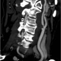
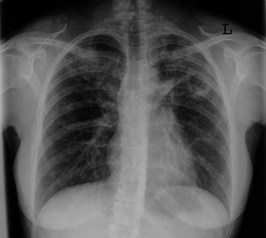
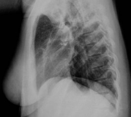
You arrange a chest X-ray. The PA and lateral views are shown in Figures 32.1 and 32.2.
Q.2
What does the chest X-ray show? What might the findings suggest? Would any other imaging be of help?
Answers
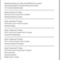
A.4 TB is the likely diagnosis, requiring the following measures:
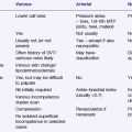
| Drug | Side-Effect | Interaction |
|---|---|---|
| Isoniazid | Peripheral neuropathy, hepatitis, rash | Anticonvulsants (increased level) |
| Rifampicin | Hepatitis, rash, flu-like syndrome, thrombocytopenia | Warfarin, oral contraceptives, oral hypoglycaemics, anticonvulsants (reduced level) |
| Pyrazinamide | Hepatitis, rash, arthralgia, gout | |
| Ethambutol* | Optic neuritis |
* Use three times weekly or avoid in those with renal impairment or avoid in those with renal impairment.
Revision Points
Pulmonary Tuberculosis
Pathology
Diagnosis
Prevention
, www.nlm.nih.gov/medlineplus/tuberculosis.html. A National Library of Medicine website. Comprehensive coverage of tuberculosis with links to a wide variety of sites dealing with many aspects of this disease
, www.who.int/gtb. A superb web resource from the World Health Organization dealing with the global fight against tuberculosis infection