Peripheral nerves

Published on 02/03/2015 by admin

Filed under Basic Science

Last modified 22/04/2025

Print this page

rate 1 star rate 2 star rate 3 star rate 4 star rate 5 star
Your rating: none, Average: 0 (0 votes)

This article have been viewed 1318 times

9 Peripheral nerves

General Features

The peripheral nerves comprise the cranial and spinal nerves linking the brain and spinal cord to the peripheral tissues. The spinal nerves are formed by the union of anterior and posterior nerve roots at their points of exit from the vertebral canal (Figure 9.1). The swelling on each posterior root is a spinal or posterior root ganglion. The spinal nerve is only 1 cm long and occupies an intervertebral foramen. On emerging from the foramen, it divides into anterior and posterior rami.

The posterior rami supply the erector spinae muscles and the overlying skin of the trunk. The anterior rami supply the muscles and skin of the side and front of the trunk, including the muscles and skin of the limbs; they also supply sensory fibers to the parietal pleura and parietal peritoneum.

The cervical, brachial, and lumbosacral plexuses are derived from anterior rami, which form the roots of the plexuses. The term root therefore has two different meanings depending on the context. (Details of the plexuses are in standard anatomy texts.)

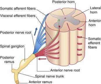
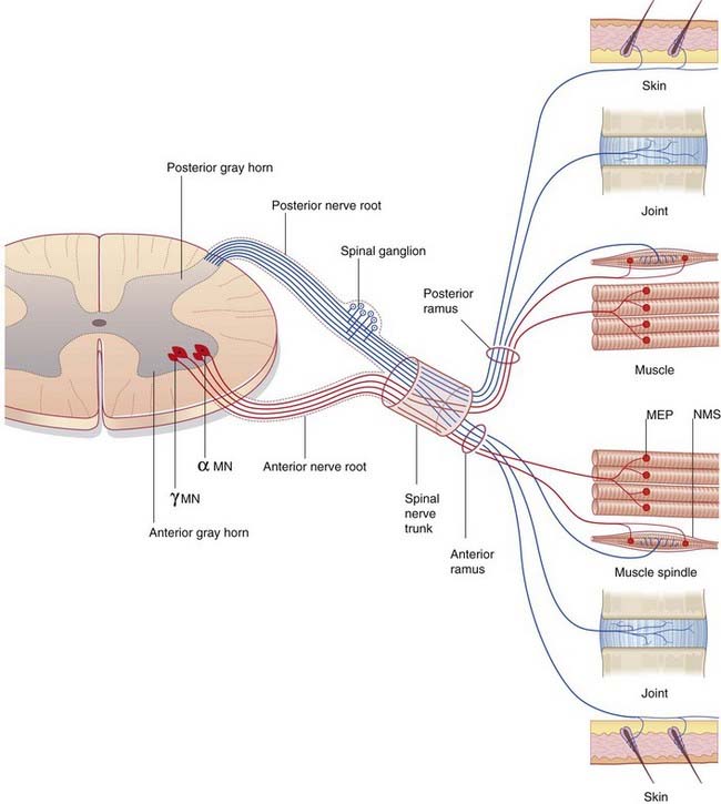
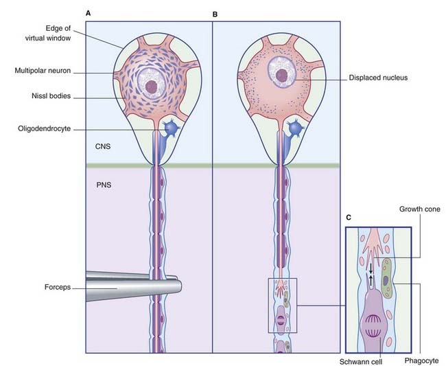
The neurons contributing to peripheral nerves are partly contained within the central nervous system (CNS) (Figure 9.2). The cells giving rise to the motor (efferent) nerves to skeletal muscles are multipolar alpha and gamma neurons of similar configuration to the one depicted in Figure 6.4; in the spinal cord, they occupy the anterior horn of gray matter. Further details are in Chapter 10. Those giving rise to posterior nerve roots are unipolar neurons whose cell bodies lie in posterior root ganglia and whose sensory (afferent) central processes enter the posterior horn of gray matter.

The spinal nerves supply somatic efferent fibers to the skeletal muscles of the trunk and limbs, and somatic afferent fibers to the skin, muscles, and joints. They all carry visceral efferent, autonomic fibers, and some spinal nerves contain visceral afferent fibers too.

Microscopic Structure of Peripheral Nerves

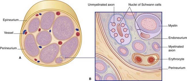
Figure 9.3 illustrates the structure of a typical peripheral nerve. It is not possible to designate individual nerve fibers as motor or sensory on the basis of structural features alone.

Peripheral nerves are invested with epineurium, a loose, vascular connective tissue sheath surrounding the fascicles (bundles of fibers) that make up the nerve. Nerve fibers are exchanged between fascicles along the course of the nerve.

Each fascicle is covered by perineurium, composed of several layers of pavement epithelium bonded by tight junctions. Surrounding the individual Schwann cells is a network of reticular collagenous fibers, the endoneurium.

Less than half of the nerve fibers are enclosed in myelin sheaths. The remaining, unmyelinated fibers travel in deep gutters along the surface of Schwann cells.

The term nerve fiber is usually used in the context of nerve impulse conduction, where it is equivalent to axon. An anatomic definition is possible for a myelinated fiber: it comprises axon, myelin, and neurolemmal sheaths, and endoneurium. It is not possible for unmyelinated axons because they share neurolemmal (Schwann cell) and endoneurial sheaths.

Myelin formation

The Schwann cell is the representative neuroglial cell of the peripheral nervous system (PNS). It forms chains of neurolemmal cells along the nerves. Modified Schwann cells form satellite cells in posterior root ganglia and in autonomic ganglia, and teloglia at encapsulated sensory nerve endings (Ch. 11).

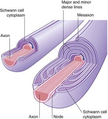
If an axon is to be myelinated, it receives the simultaneous attention of a sequence of Schwann cells along its length. Each one encloses the axon completely, creating a ‘mesentery’ of plasma membrane, the mesaxon (Figure 9.4). The mesaxon is displaced progressively, being rotated around the axon. Successive layers of plasma membrane come into apposition to form the major and minor dense lines (Figure 9.4).

Paranodal pockets of cytoplasm persist at the ends of the myelin segments, on each side of the nodes of Ranvier.

Myelin expedites conduction

Along unmyelinated fibers, impulse conduction is continuous (uninterrupted). Its maximum speed is 15 m/s. Along myelinated fibers, excitable membrane is confined to the nodes of Ranvier, because myelin is an electrical insulator. Impulse conduction is called saltatory (‘jumping’), because it leaps from node to node. Speed of conduction is much greater along myelinated fibers, with a maximum of 120 m/s. The number of impulses that can be conducted by myelinated fibers is also much greater than by unmyelinated ones.

The larger the myelinated fiber, the more rapid the conduction, because larger fibers have longer internodal segments and the nerve impulses take longer ‘strides’ between nodes. A ‘rule of six’ can be used to express the ratio between size and speed: a fiber of 10 µm external diameter (i.e. including myelin) will conduct at 60 m/s, one of 15 µm at 90 m/s, and so on.

In physiologic recordings, peripheral nerve fibers are classified in accordance with conduction velocities and other criteria. Motor fibers are classified into groups A, B, and C in descending order. Sensory fibers are classified into types I–IV. In practice, there is some interchange of usage: for example, unmyelinated sensory fibers are usually called C fibers rather than type IV.

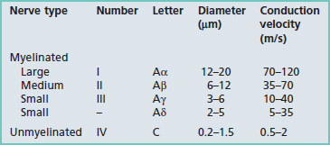
Details of diameters and sources are given in Tables 9.1 and 9.2.

Table 9.2 Locations of peripheral nerve fiber types

Fiber type Origin
Sensory  
Ia Muscle spindle annulospiral endings
Ib Golgi tendon organs
II (Aβ) Muscle spindle flower spray endings; touch or pressure receptors in skin and elsewhere
III (Aδ) Follicular endings; fast pain and thermal receptors
IV (C) Slow pain, itch, touch receptors
Motor  
Alpha motor neurons supplying extrafusal muscle fibers
Gamma motor neurons supplying intrafusal muscle fibers

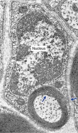
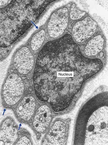
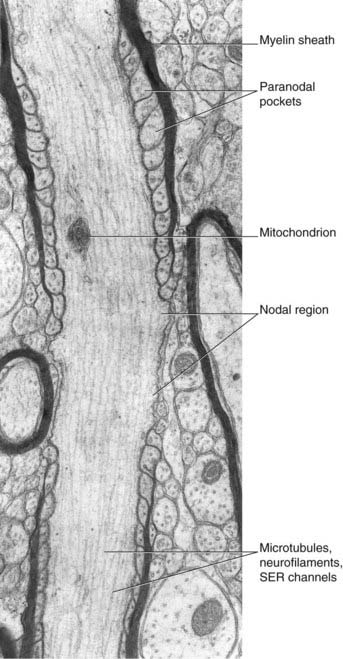
The electron micrograph in Figure 9.5 illustrates a myelinated peripheral nerve fiber with attendant Schwann cell, that in Figure 9.6 a group of unmyelinated fibers bedded in the cytoplasm of a Schwann cell, and Figure 9.7 a nodal region within the CNS.

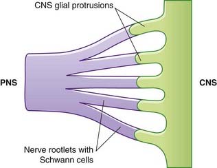
Central nervous system–peripheral nervous system transitional region

Close to the brainstem and spinal cord, peripheral nerves enter the CNS–PNS transitional zone (Figure 9.8). Astrocyte processes reach out of the CNS into the endoneurial compartments of peripheral nerve rootlets and interdigitate with the Schwann cells. In unmyelinated fibers, the astrocytes burrow into the space between axons and Schwann cells. In myelinated fibers, nodes are bounded by Schwann cell myelin (showing some transitional features) on the peripheral side, and by oligodendrocytic myelin centrally.

Degeneration and Regeneration

When nerves are cut or crushed, their axons degenerate distal to the lesion, because axons are pseudopodial outgrowths and depend on their parent cells for survival. In the PNS, regeneration is vigorous and it is often complete. In the CNS, on the other hand, it is neither vigorous nor complete.

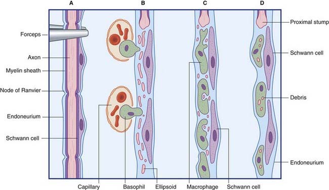
Wallerian degeneration of peripheral nerves

The principal events in peripheral nerve degeneration are represented in Figure 9.9 and described in the caption. Following a crush or cut injury to a nerve, the axons and myelin sheaths distal to the cut break up into ‘elipsoids within the first 48 h – mainly because of Ca2+-activated release of proteases by Schwann cells. The debris is cleared by monocytes that enter the damaged endoneurial sheaths from the blood and become macrophages. In addition to their phagocytic function, the macrophages are mitogenic to Schwann cells and participate with Schwann cells in provision of trophic (feeding) and tropic (guidance) factors for regenerating axons.

The end result of degeneration is a shrunken nerve skeleton with intact connective tissue and perineurial sheaths, and a core of intact, multiplying Schwann cells.

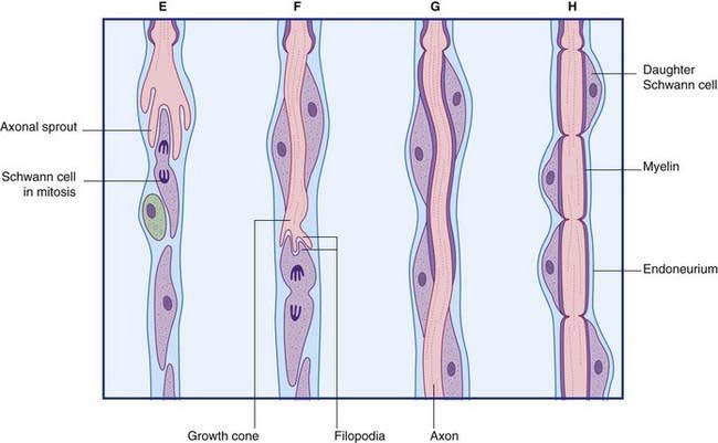
Regeneration of peripheral nerves

The principal events in regeneration of a peripheral nerve are summarized in Figure 9.9B. Following a clean cut, axons begin to sprout from the face of the proximal stump within a few hours, but in the more common crush or tear injuries seen clinically, the axons die back for 1 cm or more and sprouting may be delayed for a week. Successful regeneration requires that the axons make contact with Schwann cells of the distal stump. Failure to make contact leads to production of a pseudoneuroma consisting of whorls of regenerating axons trapped in scar tissue at the site of the initial injury. Following amputation of a limb, an amputation pseudoneuroma can be a source of severe pain.

Two reparative events are in simultaneous progress within hours of the injury. In the proximal stump, multiple branchlets begin to extend distally, their tips exhibiting swellings called growth cones; in the distal stump, Schwann cells send processes in the direction of the growth cones. The cones are surmounted by antenna-like filopodia, and these develop surface receptors that become anchored temporarily to complementary cell surface adhesion molecules in Schwann cell basement membranes. Filaments of actin within the filopodia become attached to the surface receptors; from these points of anchorage, they are able to exert onward traction on the growth cones.

Growth cones are mitogenic to Schwann cells, which divide further before wrapping the larger axons with myelin lamellae.

Regeneration proceeds at about 5 mm/day in the larger nerve trunks, slowing down to 2 mm/day in the finer branches. Not surprisingly, the functional outlook is better after a crush injury (endoneurium preserved) than after complete severance. At the same time, filopodia of motor and sensory axons ‘recognize’ Schwann cell basement membranes previously occupied by axons of similar kind.

When nerve trunks have been completely severed, it is common practice to wait about 3 weeks before attempting repair. By that time, the connective tissue sheaths will have thickened a little and will be better able to hold suture material than would freshly injured, edematous sheaths. Moreover, the trimming of the nerves required before insertion of sutures creates a second axotomy, on the axons emerging from the proximal stump. In animal experiments, a second axotomy induces a more vigorous and sustained regenerative response.

Upstream effects of nerve section are as follows:

Within a few days of axotomy, Nissl bodies can no longer be identified by cationic dyes in parent cells in the dorsal root ganglia and spinal gray matter (Figure 9.10). The phenomenon is known as chromatolysis (‘loss of color’). Electron microscopy reveals that the granular endoplasmic reticulum is in fact increased in amount. But it is now dispersed throughout the perikaryon, with accumulations located deep to the plasma membrane.

Regeneration in the central nervous system

Remarkable levels of functional recovery are often observed after CNS lesions. However, injured motor and sensory pathways do not re-establish their original connections. They regenerate for a few millimeters at most, and such synapses as developed are on other neurons close to the site of injury. Adult CNS neurons (in laboratory animals at least) do have regenerative capacity, as witnessed by their liberal sprouting and invasion of the endoneurial tubes of implanted peripheral nerves. The principal deterrents to spontaneous regeneration following CNS injury are obstruction by developing glial scar tissue, and growth inhibition by oligodendrocyte breakdown products.

One of the most active areas in neurobiological research is the use of embryonic nervous tissue to replace neurons that have been lost owing to injury or disease. The mammalian CNS in general seems to be lacking in trophic factors required for successful regeneration.

Trophic factors are also abundant in central neurons because, when these are transplanted (with immunologic precautions) into adult brain, they grow well. This approach is under investigation in animal models of Parkinson’s disease, Alzheimer’s disease, and spinal cord injury, with limited benefits in all three.