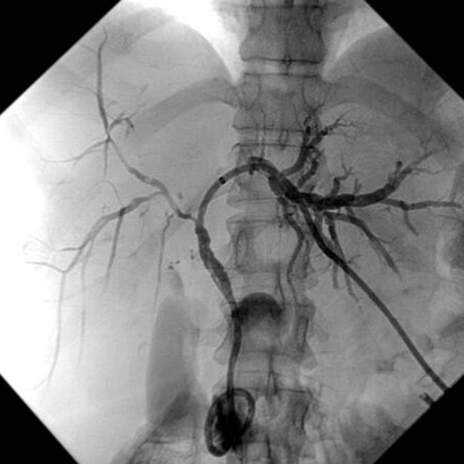
 and bile ducts
  encased and obstructed by the tumor
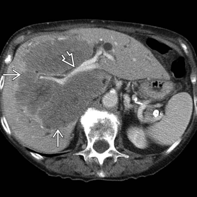
  , accounting for the altered perfusion of the right hepatic lobe. Hepatic veins were encased as well, resulting in collateral blood vessels seen within the right lobe
  .
.
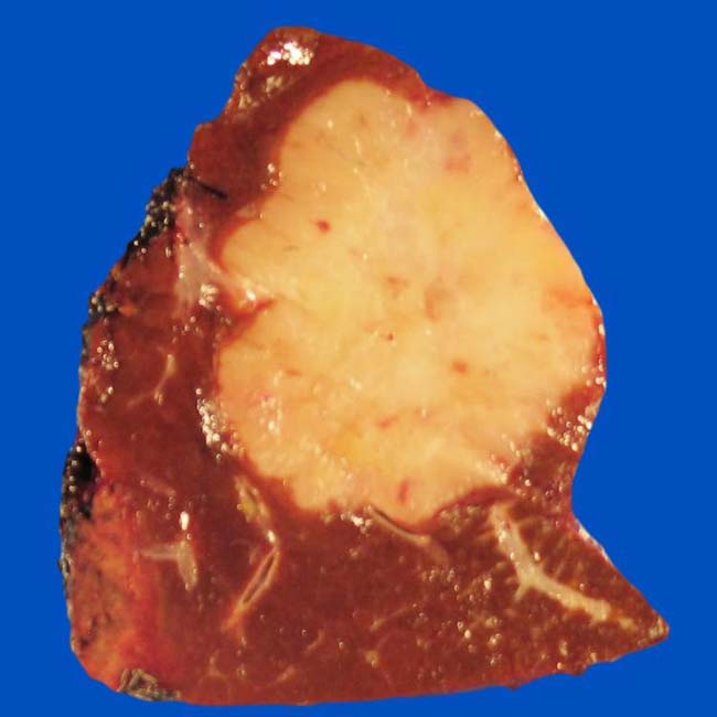
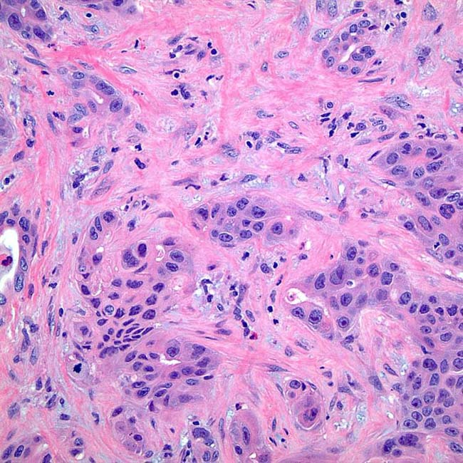
 , a feature of cholangiocarcinoma (and other tumors with fibrous stroma).
IMAGING
General Features


CT Findings

• CECT
 Mass-forming PCC
Mass-forming PCC
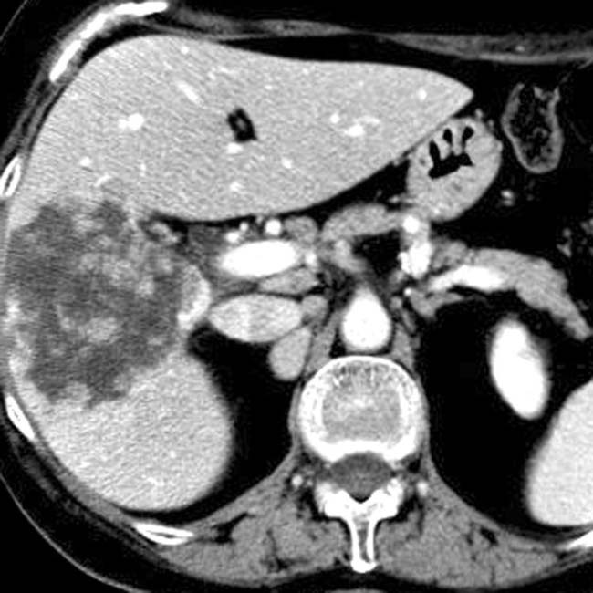
– Thin or thick rim-like enhancement frequently seen around periphery of tumor on arterial phase images


 .
.
 . The intrahepatic bile ducts are dilated
  , and the left lobe of the liver is atrophic. Parenchymal atrophy of liver segments affected by peripheral cholangiocarcinoma is common and may be evident as lobar atrophy or capsular retraction.
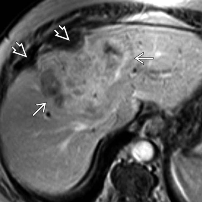
 . Capsular retraction
  is also noted. This tumor showed persistent enhancement on 10-minute delayed images.
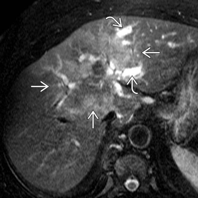
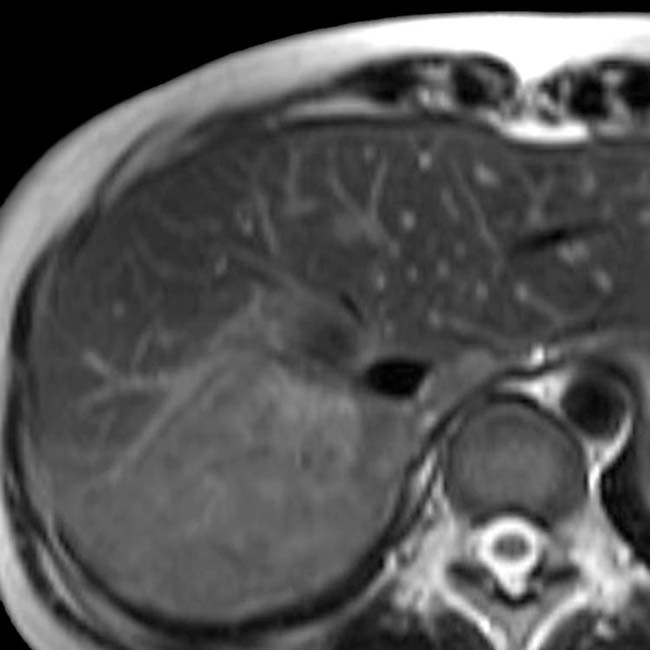
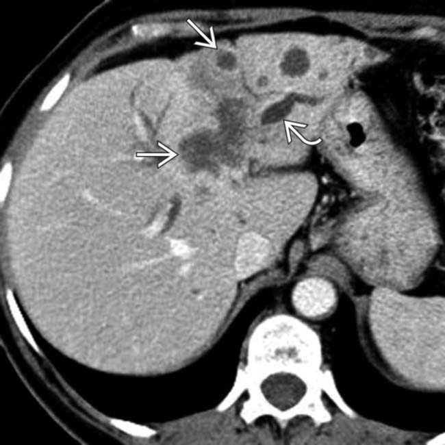
 . Dilated intrahepatic ducts
  are noted upstream from the mass. This is a typical feature of cholangiocarcinoma but may be seen with other tumors.
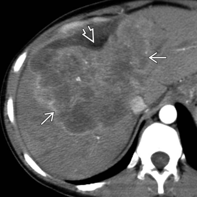
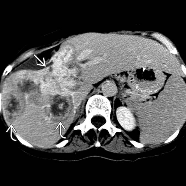
 with heterogeneous minimal enhancement. Hepatic capsular retraction
  is a prominent feature of this cholangiocarcinoma.
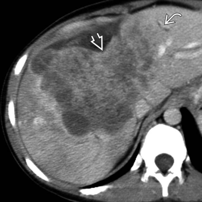
 , and there is marked retraction of the liver capsule
  .
.
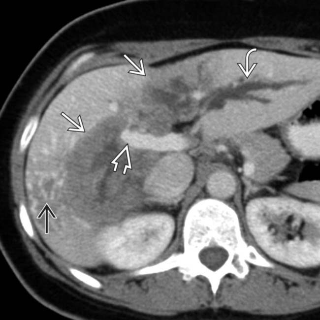
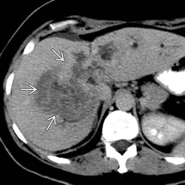
 filling much of the right lobe of the liver. The mass obstructs central bile ducts, causing dilation of the peripheral intrahepatic ducts
  .
.
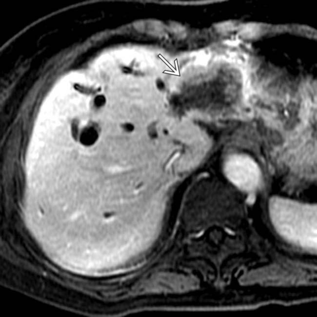
 encases the portal vein
  . The delayed set of images (not shown) showed some persistent enhancement in some parts of the tumor and no washout.
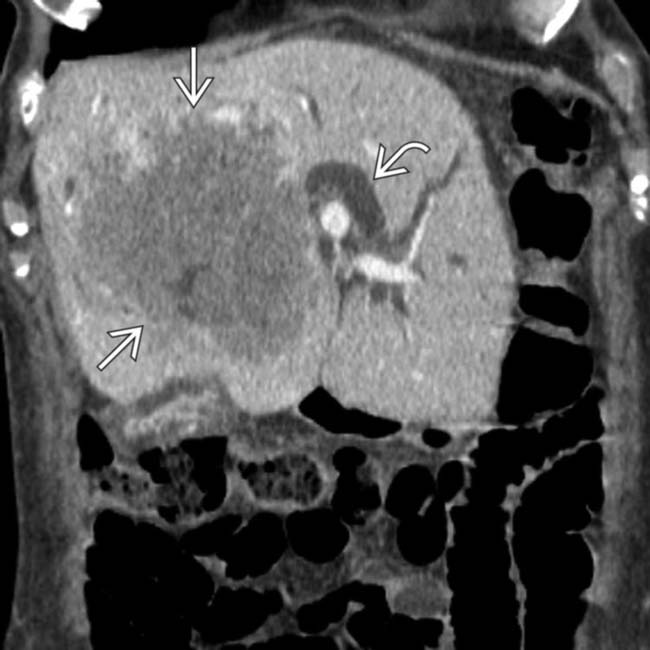
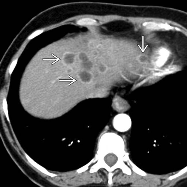
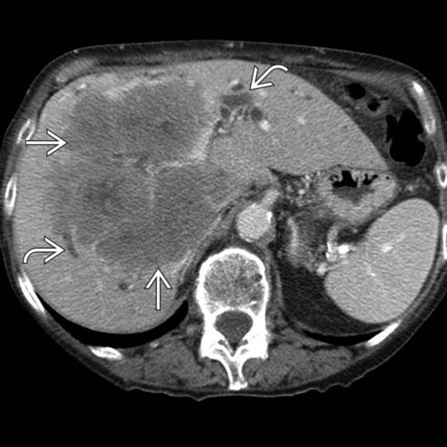
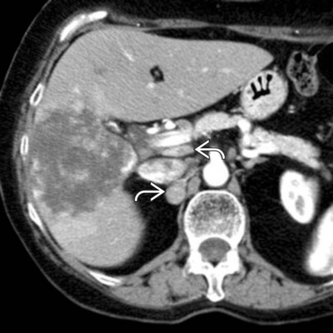
 with dilation of intrahepatic bile ducts
  . The CT findings are typical, but not diagnostic, of cholangiocarcinoma.
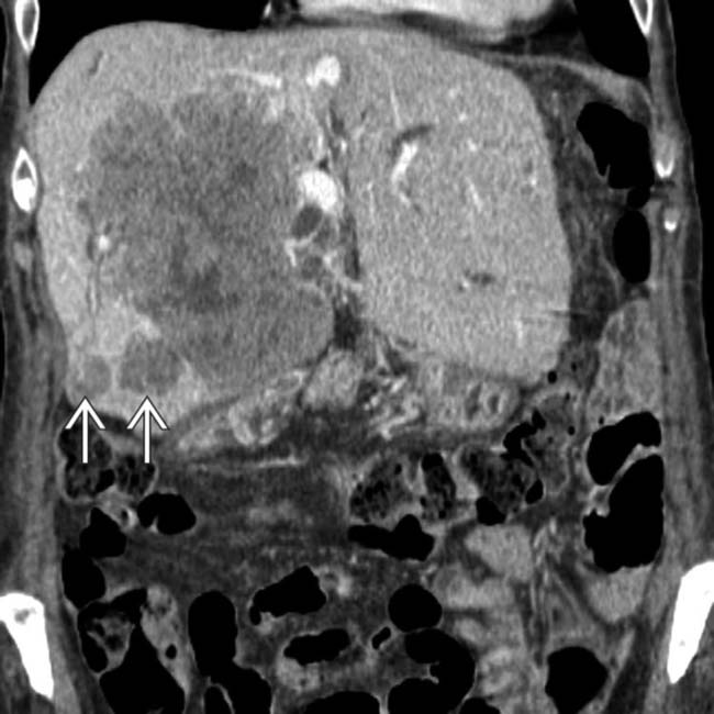
 , a common feature of intrahepatic cholangiocarcinoma. The term “peripheral” cholangiocarcinoma seems to be a misnomer in cases such as this, but it implies that the tumor arose from intrahepatic bile ducts.
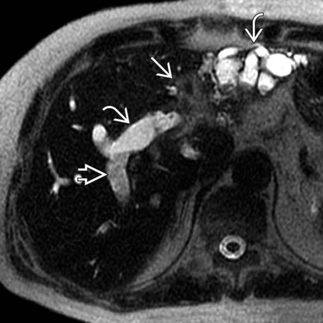
 with stones and pus within the dependent ducts
  . Marked ductal dilation plus lobar atrophy of the left lobe are caused by an obstructing mass
  .
.
 and the intrahepatic ductal dilation. Cholangiocarcinoma was confirmed at left hepatic resection.
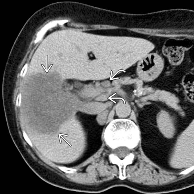
 with porta hepatic lymphadenopathy
  .
.
 have the same enhancement pattern as the intrahepatic cholangiocarcinoma, indicating nodal metastases.
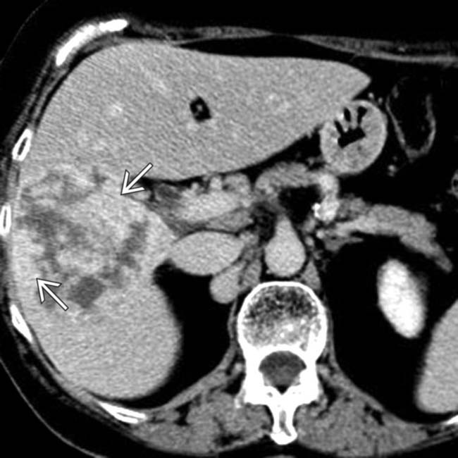
 , but the lesion does not fill in from the periphery as would be expected of a hemangioma.
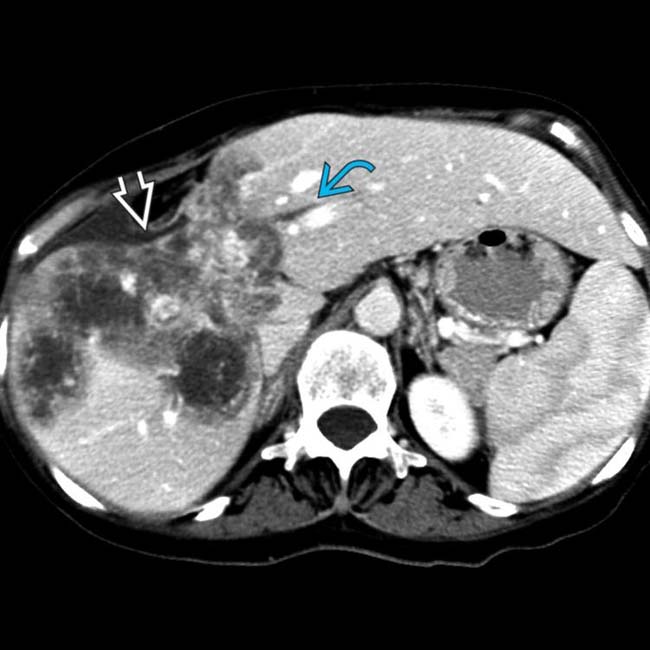
 and volume loss with capsular retraction
  .
.
 due to its fibrous stroma. Other tumor foci have the “target” pattern of enhancement
  of other types of adenocarcinomas.
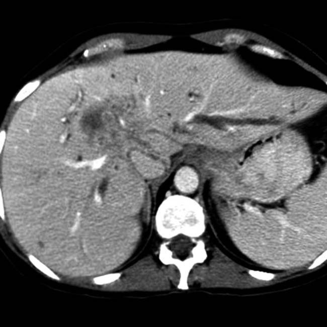
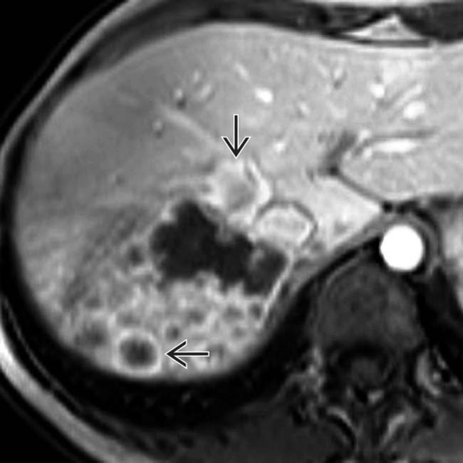
 along with low-intensity, nonenhancing (fibrotic/necrotic) areas.