[level-membership-for-pediatrics-category]
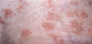
Tense blisters on the leg in childhood bullous pemphigoid.

Vulvar blisters, erosions and early scarring in a preschool child with localized vulvar pemphigoid.
CLINICAL FEATURES
Bullous pemphigoid (BP) is caused by circulating antibodies to either the 180-kd or 230-kd BP antigen (type 17 collagen). Immunofluorescence findings include linear deposition of IgG and/or C3 at the dermoepidermal junction. The infantile form of BP usually presents with blisters on the palms, soles and scalp, whereas the childhood form is more likely to present with vulvar lesions and a subset of girls with BP will have only vulvar lesions. Individual lesions vary greatly in size, are tense and filled with serous fluid that may become hemorrhagic. Lesions may be pruritic or painful and may heal with scarring. Mucosal lesions are seen less commonly than in pemphigus vulgaris. Rarer variants include those associated with lichen planus and solid organ transplant rejection.
TREATMENT
Infants and children with localized disease may be treated with super-potent topical steroids. Those with more generalized disease will require treatment with methylprednisolone (1−2 mg/kg/day). Remission is usually achieved in several weeks to a few months and relapses are very rare. There is no difference in response to treatment for any of the types of BP.
[/level-membership-for-pediatrics-category][not-level-membership-for-pediatrics-category]
Tense blisters on the leg in childhood bullous pemphigoid.
Vulvar blisters, erosions and early scarring in a preschool child with localized vulvar pemphigoid.
CLINICAL FEATURES
[/not-level-membership-for-pediatrics-category]
