[level-membership-for-orthopaedics-category]
Chapter 11 Pelvis and Lower Extremity Reduction
Pelvis reduction
Overview
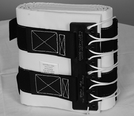
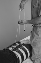
1. Application of a pelvic binder is a key step in the initial management of an unstable pelvic fracture.
Indications for Use
Precautions
Pearls
Detailed Technique
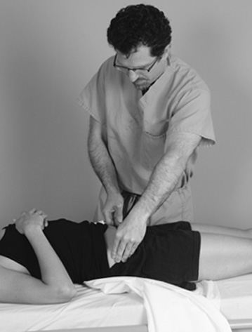
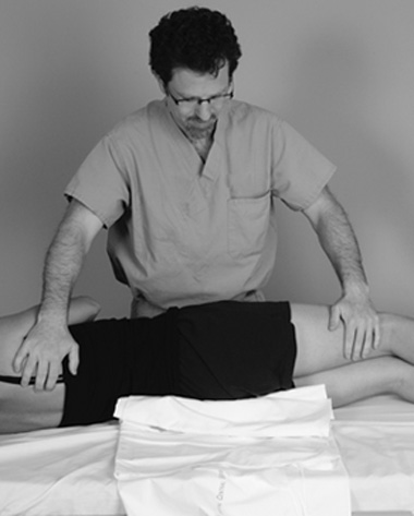
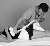
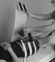
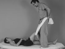
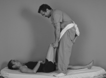
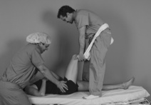
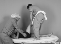
3. Roll the patient. A standard trauma log-roll technique should be used to place the commercial binder or bed sheet under the patient (Figure 11-3), with additional people at the legs and the head.
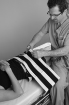
4. If a bed sheet is being used, place it as widely as possible over the greater trochanter and ASIS.
Hip reduction
Overview
3. The incidence of dislocated hips is much higher in patients with hip arthroplasty compared with patients who do not have hip arthroplasty.
5. Relocation of hips can be difficult because of the significant muscular and ligamentous impediments inherent to the joint.
Knee reduction
Overview
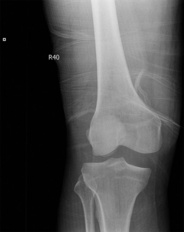
1. Dislocation of the knee is associated with a high incidence of associated vascular and nerve injuries. The popliteal artery and peroneal nerve are most commonly injured.
Precautions
4. Altered sensation in the calf and entire foot (a “stocking type” distribution) suggests a vascular injury.
Ankle fracture reduction
Overview
2. The traction reduction method is based on a modification of the reduction maneuver described by Quigley.
Indications for Use
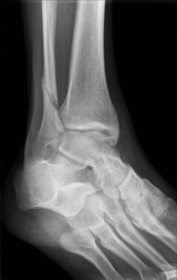
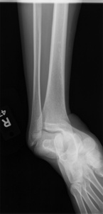
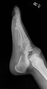
Supination external rotation ankle fractures (Figure 11-17) or fracture dislocations
Precautions
Pearls
Detailed Technique: Traction
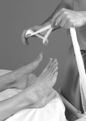
1. Set up the equipment:
3. Perform an intra-articular ankle block. Inject the ankle with lidocaine and ropivacaine (see Chapter 7).
Detailed Technique: Manual Reduction
2. Perform an intra-articular ankle block. Inject the ankle with lidocaine and ropivacaine (see Chapter 7).
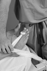
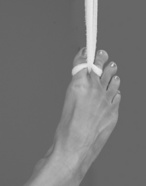
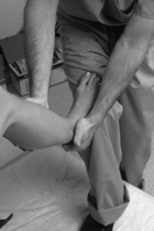
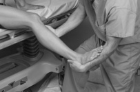
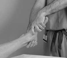
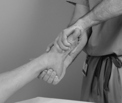
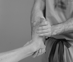
3. Cup the heel with your hand and place your other hand on the subcutaneous portion of the mid tibia while resting the ball of the foot on your thigh (Figure 11-21); alternatively, use your forearm to maintain a plantigrade foot position (Figure 11-22).
Subtalar dislocation
Overview
Precautions
Pearls
Detailed Technique
3. Perform a reduction maneuver:
a. The patient’s knee is flexed to 90 degrees and the hip is also flexed to 90 degrees by an assistant. Countertraction should be held above the ankle.
b. Medial dislocation:
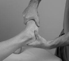
(1) The surgeon should grasp the calcaneus with his or her dominant hand and place the opposite hand over the dorsum of the forefoot (Figure 11-25).
c. Lateral dislocation:
(1) The surgeon should grasp the calcaneus with his or her dominant hand and place the opposite hand over the dorsum of the forefoot with a thumb over the navicular.
(2) The surgeon pulls, providing longitudinal traction, while an assistant provides countertraction.
[/level-membership-for-orthopaedics-category][not-level-membership-for-orthopaedics-category]
Chapter 11 Pelvis and Lower Extremity Reduction
Pelvis reduction
Overview
1. Application of a pelvic binder is a key step in the initial management of an unstable pelvic fracture.
Indications for Use
Precautions
Pearls
Detailed Technique
3. Roll the patient. A standard trauma log-roll technique should be used to place the commercial binder or bed sheet under the patient (Figure 11-3), with additional people at the legs and the head.
4. If a bed sheet is being used, place it as widely as possible over the greater trochanter and ASIS.
Hip reduction
Overview
3. The incidence of dislocated hips is much higher in patients with hip arthroplasty compared with patients who do not have hip arthroplasty.
5. Relocation of hips can be difficult because of the significant muscular and ligamentous impediments inherent to the joint.
