Pelvic Pain

Published on 10/03/2015 by admin

Filed under Obstetrics & Gynecology

Last modified 22/04/2025

Print this page

rate 1 star rate 2 star rate 3 star rate 4 star rate 5 star
Your rating: none, Average: 0 (0 votes)

This article have been viewed 3461 times

Chapter 21 Pelvic Pain

Pelvic pain is a frequent complaint in gynecology. It may be cyclic and associated with menstruation, sudden in onset (acute), or chronic, lasting for more than 6 months. Half of all menstruating women are affected by painful menstruation or dysmenorrhea, making it the most common type of pelvic pain. Ten percent of these women have severe symptoms necessitating time off from work or school.

image Dysmenorrhea

Dysmenorrhea may be primary, when there is no readily identifiable cause, or secondary to organic pelvic disease. The typical age range of occurrence for primary dysmenorrhea is between 17 and 22 years, whereas secondary dysmenorrhea is more common in older women.

PRIMARY DYSMENORRHEA

Pathophysiology

Primary dysmenorrhea occurs during ovulatory cycles and usually appears within 6 to 12 months of the menarche. The etiology of primary dysmenorrhea has been attributed to uterine contractions with ischemia and production of prostaglandins. Women with dysmenorrhea have increased uterine activity, which results in increased resting tone, increased contractility, and increased frequency of contractions. During menstruation, prostaglandins are released as a consequence of endometrial cell lysis, with instability of lysosomes and release of enzymes that break down cell membranes.

The evidence that prostaglandins are involved in primary dysmenorrhea is convincing. Menstrual fluid from women with this disorder have higher than normal levels of prostaglandins (especially prostaglandin F [PGF] and PGE2), and these levels can be reduced to below normal with nonsteroidal antiinflammatory drugs (NSAIDs), which are effective treatments. Infusions of PGF or PGE2 reproduce the discomfort and many of the associated symptoms such as nausea, vomiting, and headache. Secretory endometrium contains much more prostaglandin than proliferative endometrium. Women with primary dysmenorrhea have upregulated cyclooxygenase (COX) enzyme activity as a major cause of their pain. Anovulatory endometrium (without progesterone) contains little prostaglandin, and these menses are usually painless.

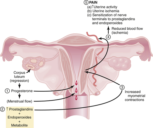
Figure 21-1 summarizes the relationships among endometrial cell wall breakdown, prostaglandin synthesis, uterine contractions, ischemia, and pain.

Treatment

Box 21-2 lists the treatment options for primary dysmenorrhea. NSAIDs, which act as COX inhibitors, are highly effective in the treatment of primary dysmenorrhea. Typical examples include ibuprofen (400 mg every 6 hours), naproxen sodium (250 mg every 6 hours), and mefenamic acid (500 mg every 8 hours). Decreasing prostaglandin production by enzyme inhibition is the basis of all NSAIDs. Hormonal contraceptives such as oral contraceptive pills (OCs), patches, or transvaginal rings reduce menstrual flow and inhibit ovulation and are also effective therapy for primary dysmenorrhea. Extended cycle use of OCs or the use of long-acting injectable or implantable hormonal contraceptives or progestin-containing intrauterine devices minimizes the number of withdrawal bleeding episodes that users have. Some patients may benefit from using both hormonal contraception and NSAIDs.

Resistant cases may respond to tocolytic agents (e.g., salbutamol), a calcium blocker (e.g., nifedipine), or high-dose continuous daily progestogens (especially medroxyprogesterone acetate or dydrogesterone). Nonpharmacologic pain management, particularly acupuncture or transcutaneous electrical stimulation, may be useful, as are psychotherapy, hypnotherapy, and heat patches. Surgical procedures such as presacral neurectomy and uterosacral ligament section have been largely abandoned.

If a patient fails to respond to hormonal contraception and NSAID therapy, the diagnosis of primary dysmenorrhea should be questioned and consideration given to a secondary cause. Ultrasonic imaging, laparoscopy, and hysteroscopy with directed biopsy should be performed to exclude pelvic disease.

SECONDARY DYSMENORRHEA

Treatment

Management consists of the treatment of the underlying disease. The treatments used for primary dysmenorrhea (Box 21-2) are often helpful. Other specific treatments are discussed in the chapters dealing with the underlying causes.

image Acute Pelvic Pain

Acute pain is sudden in onset and is usually associated with significant neuroautonomic reflexes such as nausea and vomiting, diaphoresis, and apprehension. It is important for the gynecologist to be aware of both the gynecologic and nongynecologic causes of acute pelvic pain (Box 21-4). Delay of diagnosis and treatment of acute pelvic pain increase the morbidity and even mortality.

Adnexal accidents, including torsion or rupture of an ovarian (Figure 21-2) or fallopian tube cyst, can cause severe lower abdominal pain. Normal ovaries and fallopian tubes rarely undergo torsion, but cystic or inflammatory enlargement predisposes to these adnexal accidents. The pain of adnexal torsion can be intermittent or constant, is often associated with nausea, and has been described as reverse renal colic because it originates in the pelvis and radiates to the loin. An enlarging pelvic mass is found on examination and ultrasound with decreased or absent blood flow to the adnexa on Doppler ultrasound studies. The need for surgical intervention is common and urgent.

image

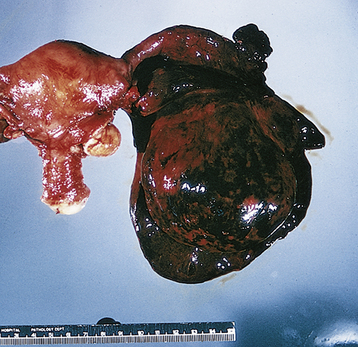
FIGURE 21-2 Torsion of an ovarian cyst.

(From Clement PB, Young RH: Atlas of Gynecologic Surgical Pathology. Philadelphia, WB Saunders, 2000.)

Functional ovarian cysts (e.g., corpus luteum or follicular cysts) may rupture, causing leakage of fluid or blood that causes acute pain from peritoneal irritation. When there is significant associated bleeding, the pain may be followed by a hemoperitoneum and hypovolemia. Surgical intervention is mandatory in this setting, after adequate resuscitation with packed red cells and intravenous fluids.

Acute reproductive organ infections such as endometritis or salpingo-oophoritis (commonly referred to as pelvic inflammatory disease [PID]) can present acutely. Rupture of a tubo-ovarian abscess is a surgical emergency that can progress to hypotension and oliguria after initially presenting with diffuse lower abdominal pain. Pelvic infection is covered in greater detail in Chapter 22.

Several complications of early pregnancy, such as ectopic gestation (see Chapter 24) and threatened or incomplete abortion can cause acute pelvic pain and are generally associated with abnormal bleeding. Ectopic tubal pregnancies produce pain as the fallopian tube dilates and ruptures into the abdominal cavity and can be life-threatening when not diagnosed expeditiously.

Nongynecologic causes of acute lower abdominal pain (see Box 21-4) are frequently seen in the differential diagnosis when a woman presents with pelvic pain. Appendicitis is a common gastrointestinal cause of acute lower abdominal pain that eventually localizes to the right lower quadrant of the abdomen (McBurney’s point). The unilateral intensity of the pain usually differentiates it from salpingo-oophoritis. Rupture of an infected appendix into the pelvic cavity can have a significant adverse effect on female fertility. Diverticular abscess is also not uncommon but usually occurs in postmenopausal women.

Acute cystitis and ureteral stone formation (lithiasis) and passage are both frequently painful. Urethral syndrome can present acutely and become chronic over time when not recognized and treated. Urinary tract disorders are covered in more detail in Chapter 23.

image Anatomy and Physiology

The pain fibers to pelvic organs are shown in Table 21-1. Painful impulses that originate in the skin, muscles, bones, joints, and parietal peritoneum travel in somatic nerve fibers, whereas those originating in the internal organs travel in visceral nerves.

TABLE 21-1 NERVES CARRYING PAINFUL IMPULSES FROM THE PELVIC ORGANS

Organ Spinal Segments Nerves
Perineum, vulva, lower vagina S2-4 Pudendal, inguinal, genitofemoral, posterofemoral cutaneous
Upper vagina, cervix, lower uterine segment, posterior urethra, bladder trigone, uterosacral and cardinal ligaments, rectosigmoid, lower ureters S2-4 Pelvic parasympathetics
Uterine fundus, proximal fallopian tubes, broad ligament, upper bladder, cecum, appendix, terminal large bowel T11-12, L1 Sympathetics through hypogastric plexus
Outer two thirds of fallopian tubes, upper ureter T9-10 Sympathetics through aortic and superior mesenteric plexus
Ovaries T9-10 Sympathetics through renal and aortic plexus and celiac and mesenteric ganglia
Abdominal wall

Visceral pain is more diffusely spread than somatic pain because of a phenomenon called viscerosomatic convergence and the lack of a well-defined projection area in the sensory cortex for its identification. Viscerosomatic convergence occurs in all second-order neurons in the dorsal horn of the spinal cord that receive visceral input. No second-order neurons in the dorsal horn receive only visceral input. The viscerosomatic neurons have larger receptive fields than do the somatic second-order neurons, and the number of somatic second-order neurons vastly exceeds the number of viscerosomatic neurons. Visceral pain is therefore usually referred to the skin, which is supplied by the corresponding spinal cord segment (referred pain). For example, the initial pain of appendicitis is referred to the epigastric area, as both structures are innervated by the thoracic cord segments T8, T9, and T10.

The structures of the female genital tract vary in their sensitivity to pain. The skin of the external genitalia is exquisitely sensitive. Pain sensation is variable in the vagina; the upper segment is somewhat less sensitive than the lower. The cervix is relatively insensitive to small biopsies but is sensitive to deep incision or to dilation. The uterus is quite sensitive. The ovaries are insensitive to many stimuli, but they are sensitive to rapid distention of the ovarian capsule or compression during physical examination.

image Patient Evaluation

HISTORY

A pain history should be obtained during the first visit. Characteristics of the pain, including its location, radiation, severity, and alleviating and aggravating factors as well as effects on menstruation, level of stress, work, exercise, and intercourse should be determined. Symptoms related to the gastrointestinal, genitourinary, and musculoskeletal and neurologic systems should be elicited. This process can be guided by the Pain History Mnemonic outlined in Box 21-5.

PHYSICAL EXAMINATION

The examination of the abdomen should be performed gently to prevent involuntary guarding, which may obstruct the findings. The patient should be asked to point to the exact location of the pain and its radiation, and an attempt should be made to duplicate the pain by palpation of each abdominal quadrant. The severity of the pain should be quantified on a 0 to 10 scale (0 = no pain, 10 = hitting thumb with a hammer).

A gentle but thorough pelvic examination should be performed with an attempt to reproduce and localize the patient’s pain. The examination may be suggestive of specific pelvic pathology. For example, patients with endometriosis may have a fixed retroverted uterus with tender uterosacral nodularity. Chronic salpingitis may be suggested by bilateral, tender, irregularly enlarged adnexal structures. A prolapsed uterus may account for pelvic pressure, pain, or low backache.

The abdominal wall should be examined for evidence of myofascial trigger points and for iliohypogastric (T12, L1), ilioinguinal (T12, L1), or genitofemoral (L1, L2) nerve entrapment. Each dermatome of the abdominal wall and back is palpated with a fingertip, and points of motion tenderness or “jump signs” are marked with a pen. The patient is asked to tense the abdominal muscles by performing a straight-leg raising maneuver (both legs raised at least 6 inches with both knees straight) or a partial sit-up. Points that are still tender or more tender or that reproduce the patient’s pain suggest that the etiology of the pain is in the abdominal wall, generally nerve entrapment, impingement, or trigger point pain, and should be injected with 2 to 3 mL of 0.25% bupivacaine. Chronic abdominal wall pain is confirmed if the pain level is reduced by at least 50% and outlasts the duration of the local anesthetic.

FURTHER INVESTIGATIONS

Psychological evaluation should be requested if an obviously traumatic event has occurred with the onset of pain; if there is obvious depression, neurosis, psychosis, or secondary gain; or to aid in the planning of pain management sessions. The latter may involve cognitive behavioral and stress reduction therapy.

Laboratory studies are of limited utility in the diagnosis of CPP, although a complete blood cell count, erythrocyte sedimentation rate (ESR), and urinalysis are indicated. The ESR is nonspecific and will be increased in any type of inflammatory condition, such as subacute salpingo-oophoritis, tuberculosis, or inflammatory bowel disease. Patients who are engaging in sexual intercourse should have a pregnancy test if they have a uterus and are not postmenopausal. Pelvic ultrasonography should be performed because the pelvic examination may miss an adnexal mass, particularly in obese patients and those who are unable to relax for the complete examination. If bowel or urinary signs and symptoms are present, an abdominal and pelvic computed tomography (CT) scan, endoscopy, cystoscopy, or CT urogram may be useful. Similarly, if there is clinical evidence of musculoskeletal disease, a lumbosacral x-ray, CT, magnetic resonance imaging, or orthopedic consultation may be in order.

Diagnostic laparoscopy is the ultimate method of diagnosis for patients with CPP of undetermined etiology. Laparoscopic examination and bimanual examination may differ in 20% to 30% of cases. Laparoscopy should only be performed if no etiology for the pain can be identified.

image Differential Diagnosis

ORGANIC CAUSES OF CHRONIC PELVIC PAIN

Of women with CPP who are subjected to diagnostic laparoscopy, about one third have no apparent pathology, one third have endometriosis, one fourth have adhesions or stigmata of chronic PID, and the remainder have other causes (Box 21-6).

UTERINE PAIN

Adenomyosis (or endometriosis interna) may cause dysmenorrhea, dyspareunia, and menorrhagia, but rarely does it cause chronic daily intermenstrual pain. Uterine myomas usually do not cause pelvic pain unless they are degenerating, undergoing torsion (twisting on their pedicles), or compressing pelvic nerves. On occasion, a submucous leiomyoma may attempt to deliver through the cervix, which may cause considerable pelvic pain akin to childbirth. Uterine myomas may cause pain from rapid growth or infarction during pregnancy.

Pelvic pain is not likely to be caused by variations in uterine position, but deep dyspareunia may occasionally be associated with uterine retroversion, especially when the uterus is fixed in place by scarring or adenomyosis. The pain has been ascribed to irritation of pelvic nerves by the stretching of the uterosacral ligaments as well as to congestion of pelvic veins secondary to retroversion. The dyspareunia is typically worse during intercourse in the missionary position and is improved in the female superior position. A tender uterus that is in a fixed retroverted position usually signifies other intraperitoneal pathology, such as endometriosis or PID, and diagnosis rests on laparoscopic findings.

PSYCHOLOGICAL FACTORS

A pathologic diagnosis may not be made in about one third of patients with CPP, even after laparoscopy, which has led to the postulation that psychological factors may be etiologic. The patients have been assumed to be anxious, neurotic, anorgasmic, and insecure in their roles as women or as mothers. When subjected to the Minnesota Multiphasic Personality Inventory (MMPI), these patients have shown a greater degree of anxiety, hypochondriasis, and hysteria than control subjects. The profiles are similar, however, in patients who have chronic pain with organic pathology, indicating that chronic pain per se engenders a complex, debilitating, psychological response. Chronic pain patients with and without pathology tend to feel depressed, helpless, and passive. They withdraw from social and sexual activity and are preoccupied with pain and suffering. Many have posttraumatic stress disorder from emotional, physical, or sexual trauma. Women with CPP are also at risk for chronic fatigue syndrome.

image Management

When treating patients with CPP, a therapeutic, supportive, and sympathetic (but structured) physician-patient relationship should be established. The patient should be given regular follow-up appointments and should not be told to call only if the pain persists. This reinforces pain behavior as a means of procuring sympathy and medical attention.

A negative evaluation or the finding of pathology not amenable to therapy (e.g., dense pelvic adhesions) does not mean that the patient should be discharged from care without therapy directed toward her symptoms. After initial reassurance that there is no serious underlying pathology, and education as to the likely mechanisms of pain production, including central nervous system factors, symptomatic therapy should be undertaken. The symptoms of pain should be approached with the seriousness and direction afforded to any other condition.

MEDICAL AND SURGICAL MANAGEMENT

The gynecologist continues to assess progress, coordinate care, and provide periodic gynecologic examinations. In the initial stages of therapy, a trial of ovulation with or without menstrual suppression with combined hormonal contraception (pills, patches, rings; cyclic or continuous), high-dose or intrauterine progestins or a gonadotropin-releasing hormone agonist may be helpful. Ovulation with or without menstrual suppression is especially helpful in patients who have midcycle, premenstrual, or menstrual exacerbation of pain and in those who have ovarian pathology, such as parovarian adhesions or recurrent functional cyst formation. NSAIDs are also useful. Pharmacologic approaches to increase inhibitory neuromodulators such as norepinephrine, serotonin (5-HT), and GABA or sodium channel blockers are frequently used in the form of tricyclic antidepressants, serotonin norepinephrine reuptake inhibitors, anticonvulsants, or other GABA-ergic agents and local anesthetics.

Surgical procedures that have not proved effective for CPP without pathology include unilateral adnexectomy for unilateral pain or total abdominal hysterectomy, presacral neurectomy, and uterine suspension for generalized pelvic pain. Lysis of adhesions is also usually nonproductive, with the possible exception of the situation in which the site of adhesions, visualized by the laparoscope, specifically coincides with the localization of pain. However, pelvic adhesions often recur after surgical lysis. Without proof of organic pathology or a reasonable functional explanation for the pelvic pain, a thorough psychosomatic evaluation should be carried out before a surgical corrective procedure is considered.

ANESTHESIA

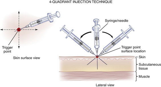
Acupuncture, nerve blocks, and trigger-point injections of local anesthetics may provide prolonged pain relief. Acupuncture has been used successfully for dysmenorrhea, and trigger-point injections and nerve blocks with local anesthetics have been used successfully for pelvic pain. Acupuncture probably increases spinal cord endorphins. In patients who complain of pelvic pain, trigger points are usually found on either the lower abdominal wall, lower back, or vaginal and vulvar areas. A significant percentage of patients with pelvic pain have abdominal wall trigger points or nerve entrapments that respond to biweekly injections of a local anesthetic (usually up to five injections is sufficient). Anesthesia of trigger points (Figure 21-3) may abolish pain by lowering the impulses from the area of referred pain, thereby diminishing the afferent impulses reaching the dorsal horn to a level below the threshold for pain transmission. Local anesthetic nerve blocks on a repeated basis for areas of nerve impingement combined with instructions to patients about alteration in physical activity can be helpful. When needed, a prescription for nerve threshold–altering medications, as mentioned previously, can be given. These interventions can downregulate neural hypersensitivity and permanently decrease or eliminate pain.

image

FIGURE 21-3 Trigger point injection technique for the abdominal wall in a patient with chronic pelvic pain.

(From Auerbach PS: Wilderness Medicine, 5th ed. Philadelphia, Mosby, 2007.)