7 Pelvic girdle
The pelvic girdle is formed by the 2 hip bones, the sacrum and the coccyx. For notes on the sacrum and coccyx, see Chapter 9.
Hip bone (Figs 7.1 and 7.2)
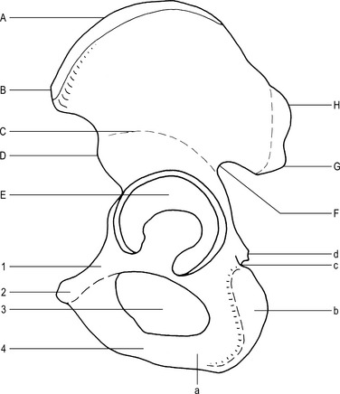
Fig. 7.1 Hip bone (external aspect).
B–Anterior superior iliac spine
D–Anterior inferior iliac spine
G–Posterior inferior iliac spine
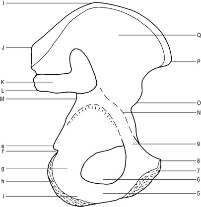
Fig. 7.2 Hip bone (internal aspect).
J–Posterior superior iliac spine
L–Posterior inferior iliac spine
P–Anterior superior iliac spine
Ilium (Figs 7.1 and 7.2)
Main parts
Posterior inferior iliac spine –
lies approximately 2.5 cm below the posterior superior iliac spine.
Posterior border –
curved border from the posterior superior iliac spine to the posterior border of the ischium.
Greater sciatic notch –
lies below the posterior inferior iliac spine. The sciatic nerve leaves the pelvis via the notch.
Sacropelvic surface –
situated between the medial and posterior borders, and divided into 3 areas:
Iliac tuberosity – upper part, roughened for ligament attachment.
Auricular surface – middle part, for articulation with the sacrum.
Pelvic surface – lower part, forms part of the wall of the true pelvis.
The pelvis (Figs 7.4 and 7.5)
The pelvis is formed posteriorly by the sacrum and coccyx and anteriorly by the two hip bones.
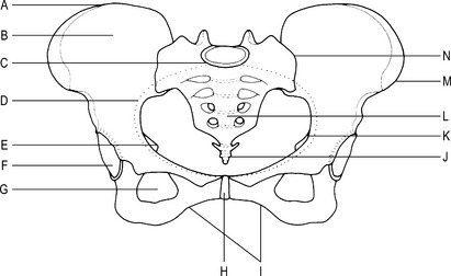
The lesser (true) pelvis
The lesser pelvis lies below the pelvic brim and can be divided into 3 areas:
The pelvic cavity
| Male | Female | |
|---|---|---|
| General shape | Narrow, deep | Wide, shallow |
| Bone structure | Heavy | Light |
| Pelvic cavity | Long, tapering downwards | Short, cylindrical |
| Sacrum | Narrow, long, curved | Wide, short, less curved |
| Pelvic inlet | Heart-shaped | Circular |
| Ischial tuberosities | Close | Wide apart |
| Pubic arch | Less than 90° | More than 90° |
| Greater sciatic notch | Acute, narrow | 90°, wide |
| Symphysis pubis | Limited movement | More flexible |
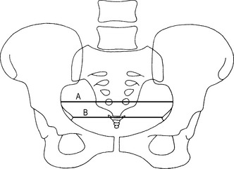
Measurement of pelvic size (Figs 7.8 and 7.9)
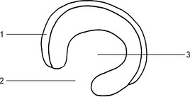
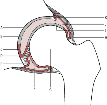
Hip joint (Figs 7.10 and 7.11)
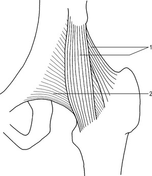
Movements
Flexion by the iliacus and the psoas assisted by the rectus femoris.
Extension by the gluteus maximus, assisted by the hamstrings.
Abduction by the gluteus medius and the gluteus minimus.
Adduction by the adductor muscles.
Lateral rotation by the obturator, gemelli and quadriceps femoris.
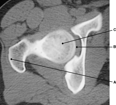
Radiographic appearances of the hip joint (Figs 7.12, 7.13 and 7.14)
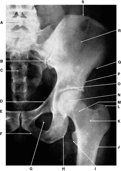
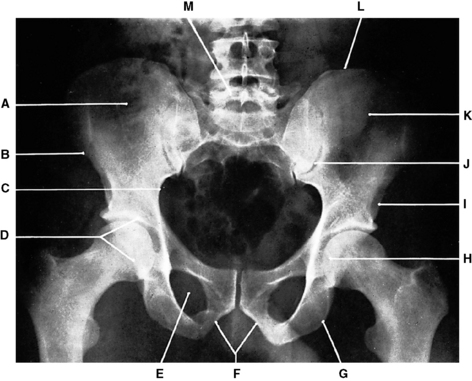
Fig. 7.12 Left hip joint: anteroposterior projection.
P–Anterior inferior iliac spine
Q–Anterior superior iliac spine
(From Bryan 1996.)
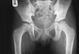
Congenital dislocation of the hip (Figs 7.15 and 7.16)
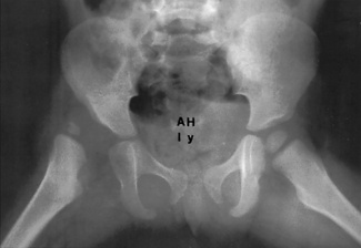
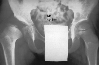
Fig. 7.15 Congenital dislocation, right hip.
a. At age 1 year. b. Same patient at age 4 years 5 months.
(Courtesy of Ernest Higginbottom.)
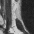
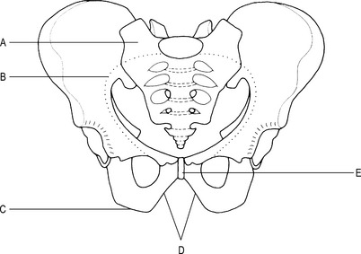
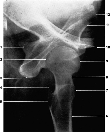
Sacroiliac joints
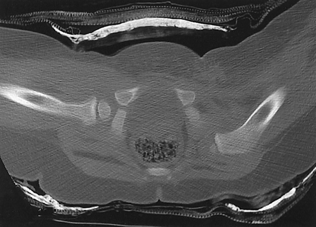
Radiographic appearances of the sacroiliac joints (Figs 7.18, 7.19, 7.20 and 7.21)
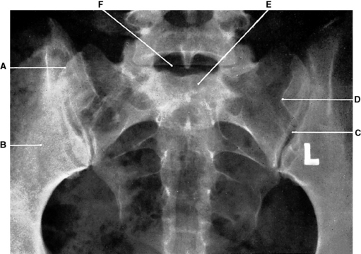
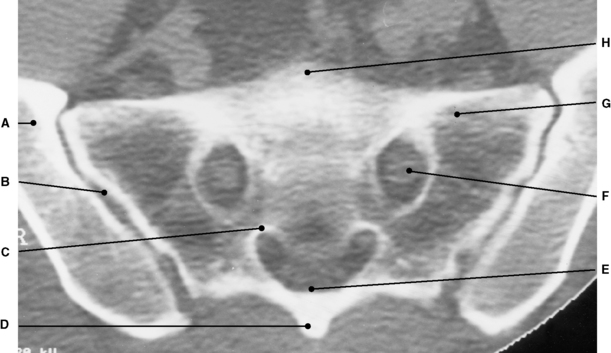
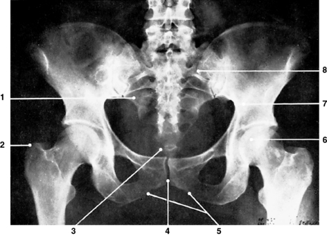
Fig. 7.18 Sacroiliac joints: anteroposterior projection.
D–Posterior superior iliac spine
(From Bryan 1996.)
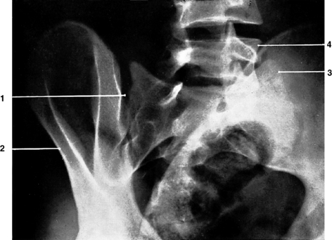
Fig. 7.19 Right sacroiliac joint: oblique projection.
4 – Body of 5th lumbar vertebra.
(From Bryan 1996.)