CHAPTER 46 Pectoral girdle, shoulder region and axilla
SKIN AND SOFT TISSUE
SKIN
Cutaneous vascular supply
The area over the lateral end of the clavicle is supplied by the supraclavicular artery which pierces the deep fascia superior to the clavicle and anterior to trapezius (Fig. 45.4). In the majority of cases this artery arises from the superficial cervical/transverse cervical artery, but it occasionally arises from the suprascapular artery. The area over the deltoid is supplied by the anterior and posterior circumflex humeral arteries. The deltoid branch of the thoraco-acromial axis contributes to the blood supply of the anterior aspect of the shoulder via musculocutaneous perforators through deltoid. For details of the cutaneous supply to the anterior, lateral and posterior chest wall see page 915.
Cutaneous innervation
The segmental supply is described in Chapter 45, page 789. The cutaneous supply to the shoulder region comes from the supraclavicular nerves (p. 436, Fig. 45.15). The floor of the axilla together with part of the upper medial aspect of the arm is supplied by the intercostobrachial nerve (lateral branch of the second intercostal nerve). Occasionally the lateral branch of the third intercostal nerve contributes to the supply of skin in the floor of the axilla. The upper lateral cutaneous nerve of the arm supplies the skin over the inferolateral part of the shoulder.
SOFT TISSUE
Deep fascia
Pectoral and axillary fascia
The pectoral fascia is a thin lamina which covers pectoralis major and extends between its fasciculi. It is attached medially to the sternum, above to the clavicle and is continuous inferolaterally with the fascia of the shoulder, axilla and thorax. Although thin over pectoralis major, it is thicker between this muscle and latissimus dorsi, and forms the floor of the axilla as the axillary fascia. The latter divides at the lateral margin of latissimus dorsi into two layers, which ensheathe the muscle and are attached behind to the spines of the thoracic vertebrae. As the fascia leaves the lower edge of pectoralis major to cross the axilla, a layer ascends under cover of the muscle and splits to envelop pectoralis minor: it becomes the clavipectoral fascia at the upper edge of pectoralis minor. The hollow of the armpit is produced mainly by the action of this fascia in tethering the skin to the floor of the axilla, and it is sometimes referred to as the suspensory ligament of the axilla. The axillary fascia is pierced by the tail of the breast (Ch. 54).
BONE
CLAVICLE
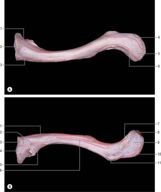
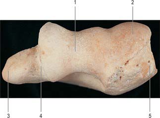
The clavicle lies almost horizontally at the root of the neck and is subcutaneous throughout its whole extent (Fig. 46.1). It acts as a prop which braces back the shoulder and enables the limb to swing clear of the trunk and transmits part of the weight of the limb to the axial skeleton. The lateral or acromial end of the bone is flattened and articulates with the medial side of the acromion, whereas the medial or sternal end is enlarged and articulates with the clavicular notch of the manubrium sterni and first costal cartilage. The shaft is gently curved and in shape resembles the italic letter f, being convex forwards in its medial two-thirds and concave forwards in its lateral third. The inferior aspect of the intermediate third is grooved in its long axis. The clavicle is trabecular internally, with a shell of much thicker compact bone in its shaft. Although elongated, the clavicle is unlike typical long bones in that it usually has no medullary cavity.
The female clavicle is shorter, thinner, less curved and smoother, and its acromial end is carried lower than the sternal in comparison with the male. In males the acromial end is on a level with, or slightly higher than, the sternal end when the arm is pendent. Midshaft circumference is the most reliable single indicator of sex: a combination of this measurement with weight and length yields better results. The clavicle is thicker and more curved in manual workers, and its ridges for muscular attachment are better marked.
Lateral third
The posterior border, also roughened by muscular attachments, is convex backwards. The superior surface is roughened near its margins but is smooth centrally, where it can be felt through the skin. The inferior surface presents two obvious markings. Close to the posterior border, at the junction of the lateral fourth with the rest of the bone, there is a prominent conoid tubercle which gives attachment to the conoid part of the coracoclavicular ligament. A narrow, roughened strip, the trapezoid line, runs forwards and laterally from the lateral side of this tubercle, almost as far as the acromial end (Fig. 46.1B). The trapezoid part of the coracoclavicular ligament is attached to it. A small oval articular facet, for articulation with the medial aspect of the acromion, faces laterally and slightly downwards at the lateral end of the shaft.
Subclavius lies in a groove on the inferior surface (Fig. 46.1B). The clavipectoral fascia is attached to the edges of the groove; the posterior edge of the groove runs to the conoid tubercle, where fascia and conoid ligament merge. Lateral to the groove there is a laterally inclined nutrient foramen. Deltoid (anterior) and trapezius (posterior) are attached to the lateral third of the shaft: both muscles reach the superior surface. The coracoclavicular ligament, which is attached to the conoid tubercle and trapezoid line (Fig. 46.1B), transmits the weight of the upper limb to the clavicle, counteracted by trapezius which supports its lateral part. From the conoid tubercle this weight is transmitted through the medial two-thirds of the shaft to the axial skeleton.
Medial two-thirds
The medial two-thirds provide attachment, anteriorly, for the clavicular head of pectoralis major: the area is usually clearly indicated on the bone. The clavicular head of sternocleidomastoid is attached to the medial half of the superior surface, but the marking on the bone is not very conspicuous. The smooth, posterior surface is devoid of muscular attachments except at its lower part immediately adjoining the sternal end, where the lateral fibres of sternohyoid are attached. Medially, this surface is related to the lower end of the internal jugular vein (from which it is separated by sternohyoid), the termination of the subclavian vein, and the start of the brachiocephalic vein. More laterally, it arches in front of the trunks of the brachial plexus and the third part of the subclavian artery. The suprascapular vessels are related to the upper part of this surface. The inferior surface gives insertion to subclavius in the subclavian groove: the clavipectoral fascia, which encloses subclavius, is attached to the edges of the groove. The posterior lip of the groove runs into the conoid tubercle and carries the fascia into continuity with the conoid ligament. A nutrient foramen is found in the lateral end of the groove, running in a lateral direction: the nutrient artery is derived from the suprascapular artery. The impression for the costoclavicular ligament is very variable in its character.
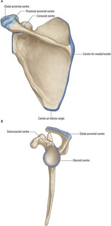
Ossification
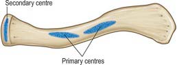
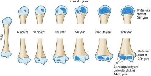
The clavicle begins to ossify before any other bone in the body, and is ossified from three centres. The shaft of the bone is ossified in condensed mesenchyme from two primary centres, medial and lateral, which appear between the fifth and sixth weeks of intrauterine life, and fuse about the 45th day. Cartilage then develops at both ends of the clavicle. The medial cartilaginous mass contributes more to growth in length than does the lateral mass: the two centres of ossification meet between the middle and lateral thirds of the clavicle. A secondary centre for the sternal end appears in late teens, or even early twenties, usually 2 years earlier in females (Fig. 46.2). Fusion is probably rapid but reliable data are lacking. An acromial secondary centre sometimes develops at around 18 to 20 years, but this epiphysis is always small and rudimentary and rapidly joins the shaft.
SCAPULA
The scapula is a large, flat, triangular bone which lies on the posterolateral aspect of the chest wall, covering parts of the second to seventh ribs (Fig. 46.3, Fig. 46.4). It has costal and dorsal surfaces, superior, lateral and medial borders, inferior, superior and lateral angles, and three processes, the spine, its continuation the acromion and the coracoid process. The lateral angle is truncated and bears the glenoid cavity for articulation with the head of the humerus. This part of the bone may be regarded as the head, and it is connected to the plate-like body by an inconspicuous neck. The long axis of the scapula is nearly vertical and the relatively featureless costal surface can easily be distinguished from the dorsal surface, which is interrupted by the shelf-like projection of the spine (Fig. 46.3A). The bone is very much thickened in the immediate neighbourhood of the lateral border, which runs from the inferior angle below, to the glenoid cavity above. The main processes, and thicker parts of the scapula, contain trabecular bone; the rest consists of a thin layer of compact bone. The central supraspinous fossa and the greater part of the infraspinous fossa are thin and even translucent; occasionally the bone in them is deficient, and the gaps are filled by fibrous tissue.
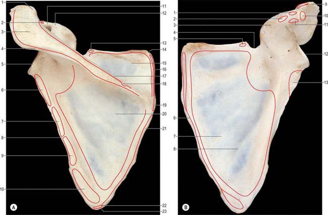
Costal surface
The costal surface, which is directed medially and forwards when the arm is by the side, is slightly hollowed out, especially in its upper part (Fig. 46.3B).
Dorsal surface
The dorsal surface is divided by the shelf-like spine of the scapula into a small upper and a large lower area, which are confluent at the spinoglenoid notch between the lateral border of the spine and the dorsal aspect of the neck (Fig. 46.3A).
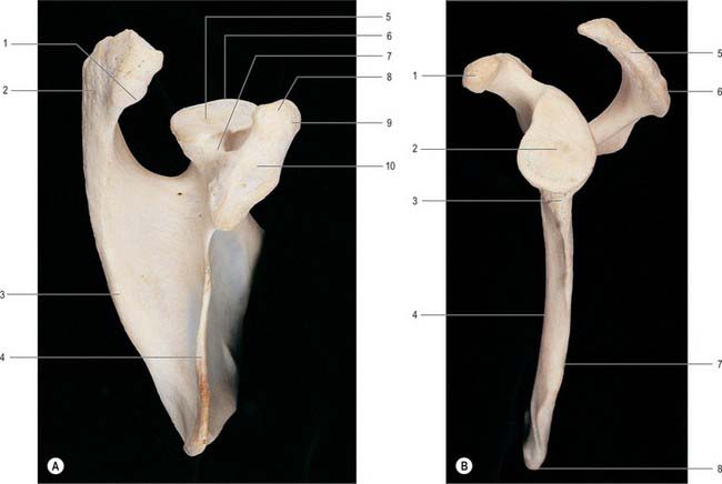
Superior border
The superior border, thin and sharp, is the shortest. At its anterolateral end it is separated from the root of the coracoid process by the suprascapular notch (Fig. 46.3B). Near the suprascapular notch it gives origin to the inferior belly of omohyoid. The notch is bridged by the superior transverse ligament which is attached laterally to the root of the coracoid process and medially to the limit of the notch. The ligament is sometimes ossified. The foramen, thus completed, transmits the suprascapular nerve to the supraspinous fossa, whereas the suprascapular vessels pass backwards above the ligament.
Lateral border
The lateral border of the scapula forms a clearly defined, sharp, roughened ridge, which runs sinuously from the inferior angle to the glenoid cavity. At its upper end it widens into a rough, somewhat triangular area, which is termed the infraglenoid tubercle (Fig. 46.4B). The lateral border separates the attachments of subscapularis and teres minor and major. These muscles project beyond the bone and, with latissimus dorsi, cover it so completely that it cannot be felt through the skin. The long head of triceps is attached to the infraglenoid tubercle.
The grooved part of the costal surface, the narrow flat lateral strip of the dorsal surface and the adjacent thickened ridge (Fig. 46.4B), are often included in the ‘lateral border’ during clinical examination.
Scapular angles
The inferior angle overlies the seventh rib or intercostal space. Palpable through the skin and covering muscles, it is also visible as it advances round the thoracic wall when the arm is raised. It is covered on its dorsal aspect by the upper border of latissimus dorsi, a small slip from which is frequently attached to the inferior angle. The superior angle, at the junction of the superior and medial borders, is obscured by the upper part of trapezius. The lateral angle, truncated and broad, bears the glenoid cavity which articulates with the head of the humerus at the glenohumeral joint. It provides a shallow, and limited, socket for the humeral head. Its outline is piriform, narrower above (Fig. 46.4B). The glenohumeral ligaments are attached to its anterior margin. When the arm is by the side, the cavity is directed forwards, laterally and slightly upwards. When the arm is raised above the head it is directed almost straight upwards. Just above it a small rough supraglenoid tubercle encroaches on the root of the coracoid process. The anatomical neck, the constriction adjoining the rim of the glenoid cavity, is most distinct at its inferior and dorsal aspects. Anteriorly and posteriorly it extends between the infraglenoid and supraglenoid tubercles, passing lateral to the root of the coracoid process. The long head of biceps brachii is attached to the supraglenoid tubercle, and the long head of triceps brachii is attached to the infraglenoid tubercle.
Spine of the scapula
The spine of the scapula forms a shelf-like projection on the upper part of the dorsal surface of the bone, and is triangular in shape (Fig. 46.3A). Its lateral border is free, thick and rounded and helps to bound the spinoglenoid notch, which lies between it and the dorsal surface of the neck of the bone. Its anterior border joins the dorsal surface of the scapula along a line which runs laterally and slightly upwards from the junction of the upper and middle thirds of the medial border. The plate-like body of the bone is bent along this line, which accounts for the concavity of the upper part of the costal surface. The dorsal border is the crest of the spine, and is subcutaneous throughout nearly its whole extent. At its medial end the crest expands into a smooth, triangular area. Elsewhere the upper and lower edges and the surface of the crest are roughened for muscular attachments. The upper surface of the spine widens as it is traced laterally, and is slightly hollowed out. Together with the upper area of the dorsal surface of the bone, the upper surface of the spine forms the supraspinous fossa. The lower surface is overhung by the crest at its medial, narrow end, but is gently convex in its wider, lateral portion. Together with the lower area of the dorsal surface of the bone, the lower surface of the spine forms the infraspinous fossa, which communicates with the supraspinous fossa through the spinoglenoid notch.
Coracoid process
The coracoid process arises from the upper border of the head of the scapula and is bent sharply so as to project forwards and slightly laterally (Fig. 46.3B, Fig. 46.4A). When the arm is by the side, the coracoid process points almost straight forwards and its slightly enlarged tip can be felt through the skin, although it is covered by the anterior fibres of deltoid. The supraglenoid tubercle marks the root of the coracoid process where it adjoins the upper part of the glenoid cavity. There is another impression on the dorsal aspect of the coracoid process at the point where it changes direction: it gives attachment to the conoid part of the coracoclavicular ligament.
Ligaments
The main scapular ligaments are the coracoacromial and superior transverse scapular; there may also be a weaker, variable inferior transverse scapular (spinoglenoid) ligament (see below Fig. 46.13, see below Fig. 46.14).
Ossification
The cartilaginous scapula is ossified from eight or more centres: one in the body, two each in the coracoid process and the acromion, one each in the medial border, inferior angle and lower part of the rim of the glenoid cavity (Fig. 46.5). The centre for the body appears in the eighth intrauterine week. Ossification begins in the middle of the coracoid process in the first year or in a small proportion of individuals before birth and the process joins the rest of the bone about the 15th year. At or soon after puberty centres of ossification occur in the rest of the coracoid process (subcoracoid centre), in the rim of the lower part of the glenoid cavity, frequently at the tip of the coracoid process, in the acromion, in the inferior angle and contiguous part of the medial border and in the medial border. A variable area of the upper part of the glenoid cavity, usually the upper third, is ossified from the subcoracoid centre; it unites with the rest of the bone in the 14th year in the female and the 17th year in the male. A horse-shoe shaped epiphysis appears for the rim of the lower part of the glenoid cavity; thicker at its peripheral than at its central margin, it converts the flat glenoid cavity of the child into the gently concave fossa of the adult. The base of the acromion is formed by an extension from the spine; the rest of the acromion is ossified from two centres which unite and then join the extension from the spine. The various epiphyses of the scapula have all joined the bone by about the 20th year.
HUMERUS
The humerus, the longest and largest bone in the upper limb, has expanded ends and a shaft (Fig. 46.6, Fig. 46.7, Fig. 46.8). The rounded head occupies the proximal and medial part of the upper end of the bone and forms an enarthrodial articulation with the glenoid cavity of the scapula. The lesser tubercle projects from the front of the shaft, close to the head, and is limited on its lateral side by a well-marked groove. The distal end, loosely termed ‘condylar’, is adapted to the forearm bones at the elbow joint.
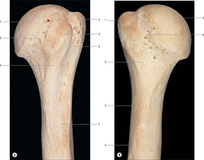
Proximal end
The proximal end of the humerus consists of the head, anatomical neck and the greater and lesser tubercles. It joins the shaft at an ill-defined ‘surgical neck’, which is closely related on its medial side to the axillary nerve and posterior humeral circumflex artery (Fig. 46.7).
Head
The head of the humerus forms rather less than half a spheroid; in sectional profile it is spheroidal (strictly ovoidal) (Fig. 46.7). Its smooth articular surface is covered with hyaline cartilage, which is thicker centrally. When the arm is at rest by the side, it is directed medially, backwards and upwards to articulate with the glenoid cavity of the scapula. The humeral articular surface is much more extensive than the glenoid cavity, and only a portion of it is in contact with the cavity in any one position of the arm.
Anatomical neck
The anatomical neck of the humerus immediately adjoins the margin of the head and forms a slight constriction, which is least apparent in the neighbourhood of the greater tubercle, and superiorly, and for some distance anteriorly and posteriorly: it indicates the line of capsular attachment of the shoulder joint (Fig. 46.7), other than at the intertubercular sulcus, where the long tendon of biceps emerges. Medially, the capsular attachment diverges from the anatomical neck and descends 1 cm or more onto the shaft.
Lesser tubercle
The lesser tubercle is anterior to and just beyond the anatomical neck. It has a smooth, muscular impression on its upper part palpable through the thickness of deltoid 3 cm below the acromial apex. The bony prominence slips away from the examining finger when the humerus is rotated. The lateral edge of the lesser tubercle is sharp and forms the medial border of the intertubercular sulcus. Subscapularis is attached to the lesser tubercle (Fig. 46.6A), and the transverse ligament of the shoulder joint is attached to the lateral margin.
Greater tubercle
The greater tubercle is the most lateral part of the proximal end of the humerus. It projects beyond the lateral border of the acromion. Its posterosuperior aspect, near the anatomical neck, bears three smooth flattened impressions for the attachment of supraspinatus (uppermost), infraspinatus (middle) and teres minor (lowest and placed on the posterior surface of the tubercle) (Fig. 46.6). The attachments of subscapularis and teres minor are not confined to their respective tubercles, but extend for varying distances on to the adjacent metaphysis. The projecting lateral surface of the tubercle presents numerous vascular foramina and is covered by the thick, fleshy deltoid, which produces the normal rounded contour of the shoulder. A part of the subacromial bursa may cover the upper part of this area and separate it from deltoid.
Shaft
Surfaces
The anterolateral surface is bounded by the anterior and lateral borders and is smooth and featureless in its upper part, which is covered by deltoid. About, or a little above, the middle of this surface, deltoid is inserted into the deltoid tubercle; further distally the surface gives origin to the lateral fibres of brachialis, which extend upwards into the floor of the lower end of the groove for the radial nerve (Fig. 46.6). Brachioradialis is attached to the proximal two-thirds of the roughened anterior aspect of the lateral supracondylar ridge, and extensor carpi radialis longus is attached to its distal third. Behind these muscles, the ridge gives attachment to the lateral intermuscular septum of the arm.
The anteromedial surface is bounded by the anterior and medial borders. Rather less than its upper third forms the rough floor of the intertubercular sulcus; the rest of the surface is smooth. Distal to the intertubercular sulcus a small area of the anteromedial surface is devoid of muscular attachment, but its lower half is occupied by the medial part of brachialis (Fig. 46.6A). Coracobrachialis is attached to a roughened strip on the middle of the medial border. The humeral head of pronator teres is attached to a narrow area close to the lowest part of the medial supracondylar ridge, and the ridge itself gives attachment to the medial intermuscular septum of the arm.
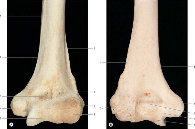
Distal end
The distal end of the humerus is a modified condyle: it is wider transversely and has articular and non-articular parts (Fig. 46.8, Fig. 46.9). The articular part is curved forwards, so that its anterior and posterior surfaces lie in front of the corresponding surfaces of the shaft. It articulates with the radius and the ulna at the elbow joint, and is divided by a faint groove into a lateral capitulum, and a medial trochlea.
Lateral epicondyle
The lateral border of the humerus terminates at the lateral epicondyle, and its lower portion constitutes the lateral supracondylar ridge. The lateral epicondyle occupies the lateral part of the non-articular portion of the condyle, but does not project beyond the lateral supracondylar ridge. It turns slightly forwards, unlike the medial epicondyle, which turns slightly backwards. Its lateral and anterior surfaces show a well-marked impression for the superficial group of the extensor muscles of the forearm (Fig. 46.6B), which arise from the lateral side of the lower humeral epiphysis, and, like the flexors, are situated outside the articular capsule. The posterior surface, which is very slightly convex, is easily felt in a depression visible behind the extended elbow. A small area on the posterior surface gives origin to anconeus.
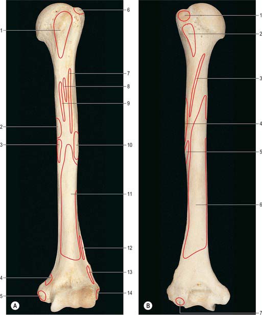
Ossification
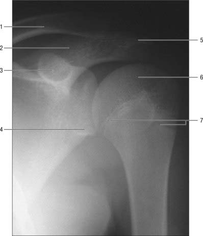
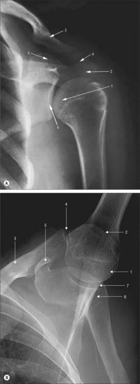
The humerus is ossified from eight centres, in the shaft, head, greater and lesser tubercles, capitulum with the lateral part of the trochlea, the medial part of the trochlea, and one for each epicondyle (Fig. 46.10). The centre for the shaft appears near its middle in the eighth week of intrauterine life, and gradually extends towards the ends. Before birth (20%), or in the first six months afterwards, ossification begins in the head, during the first year in females and second year in males in the greater tubercle, and about the fifth in the lesser tubercle. The existence of a centre in the lesser tubercle is often questioned, perhaps because it is often obscured in the usual anteroposterior radiological views (Fig. 46.11).
JOINTS
STERNOCLAVICULAR JOINT
The articulating surfaces are the sternal end of the clavicle and the clavicular notch of the sternum, together with the adjacent superior surface of the first costal cartilage (Fig. 46.12). The larger clavicular articular surface is covered by fibrocartilage, which is thicker than the fibrocartilaginous lamina on the sternum. The joint is convex vertically but slightly concave anteroposteriorly, and is therefore sellar; the clavicular notch of the sternum is reciprocally curved, but the two surfaces are not fully congruent. An articular disc completely divides the joint.
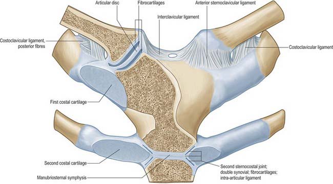
The costoclavicular ligament is like an inverted cone, but short and flattened. It has anterior and posterior laminae which are attached to the upper surface of the first rib and costal cartilage, and ascends to the margins of an impression on the inferior clavicular surface at its medial end. Fibres of the anterior lamina ascend laterally and those of the posterior lamina (which are shorter) ascend medially (Fig. 46.12); they fuse laterally and merge medially with the capsule.
ACROMIOCLAVICULAR JOINT
The acromioclavicular joint is a synovial plane joint.
The articulating surfaces are between the acromial end of the clavicle and the medial acromial margin (Fig. 46.13). The joint is approximately plane, but either surface may be slightly convex, the other being reciprocally concave, and both are covered by fibrocartilage. The clavicular surface is a narrow, oval area which faces inferolaterally and overlaps a corresponding facet on the medial acromial border. The long axis is anteroposterior.
The coracoclavicular ligament connects the clavicle and the coracoid process of the scapula (Fig. 46.14). Though separate from the acromioclavicular joint, it is a most efficient accessory ligament, and maintains the apposition of the clavicle to the acromion. The trapezoid and conoid parts of the ligament, usually separated by fat or, frequently, by a bursa, connect the medial horizontal part of the coracoid process and lateral end of the subclavian groove of the clavicle; these adjacent areas may even be covered by cartilage to form a coracoclavicular joint.
The trapezoid part is anterolateral and is broad, thin and quadrilateral, ascending slightly from the upper coracoid surface to the trapezoid line on the inferior clavicular surface. It is almost horizontal, its anterior border is free, and its posterior border is joined to the conoid part, forming an angle which projects backwards.
MOVEMENTS OF THE PECTORAL (SHOULDER) GIRDLE
GLENOHUMERAL (SHOULDER) JOINT
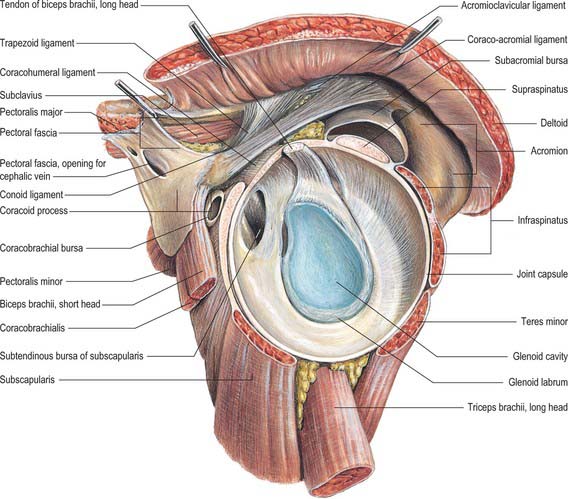
The glenohumeral joint is a synovial multiaxial spheroidal joint between the roughly hemispherical head of the humerus and the shallow glenoid fossa of the scapula (Fig. 46.15). Notable for its relative lack of bony constraint, the joint possesses three degrees of freedom. Its static and dynamic stability depends on the surrounding muscular and soft tissue envelope more than on its shape and ligaments: effective function is achieved by a complex interaction between the articular and soft tissue restraints. It is the most mobile joint in the body and the most frequently dislocated.
The articular surfaces are reciprocally curved and are really ovoids. (Here, as in the hip, where ovoid surfaces are almost spherical they are often termed spheroidal.) The area of the humeral convexity exceeds that of the glenoid concavity such that only a small portion opposes the glenoid in any position (Fig. 46.16). The remaining capitular articular surface is in contact with the capsule, so that contact on the glenoid fossa is much more uniformly distributed over its entire articular surface. The radius of curvature of the glenoid fossa in the coronal plane is greater than that of the humeral head, and is deepened by a fibrocartilaginous rim, the glenoid labrum (Fig. 46.13, Fig. 46.17). Both articular surfaces are covered by hyaline cartilage, which is thickest centrally and thinner peripherally over the humerus, and the reverse in the glenoid cavity. In most positions, their curvatures are not fully congruent, and the joint is loose-packed. Close packing (full congruence) is reached with the humerus abducted and laterally rotated (Fig. 5.61).
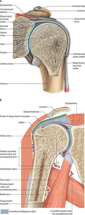
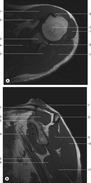
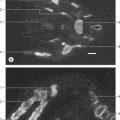
Fig. 46.17 MRI of shoulder. A, Axial image. B, Median sagittal oblique image at base of coracoid. A: 1. Deltoid. 2. Coracobrachialis. 3. Glenoid. 4. Subscapularis. 5. Infraspinatus. 6. Tendon of the long head of biceps. 7. Head of humerus. 8. Posterior labrum. B: 1. Clavicle. 2. Supraspinatus. 3. Coracoid. 4. Subscapularis. 5. Axillary nerve. 6. Latissimus dorsi. 7. Trapezius. 8. Scapular spine. 9. Infraspinatus. 10. Deltoid. 11. Triceps. Compare A with Fig. 46.23.
A fibrous capsule envelops the joint (Fig. 46.14, Fig. 46.16). It is attached medially to the glenoid neck outside the glenoid labrum, and encroaches on the coracoid process to include the attachment of the long head of biceps. The capsule often extends and attaches to the base of the coracoid and to the body of the scapula, forming anterior and posterior recesses. Laterally, it is attached to the anatomical neck of the humerus, i.e. near the articular margin, except inferomedially, where it descends more than 1 cm on the humeral shaft. It is so lax that the bones can be distracted for 2 or 3 cm, which accords with the very wide range of movement possible at the glenohumeral joint. However, such unnatural separation requires relaxation of the upper capsule by abduction.
The fibrous capsule is supported by the tendons of supraspinatus (above), infraspinatus and teres minor (behind), subscapularis (in front) and by the long head of triceps (below). The rotator interval is a medially based triangular area of uncovered capsule between the superior edge of subscapularis and the anterior edge of supraspinatus as these tendons pass on either side of the base of the coracoid: it may represent an area of weakness that increases instability in some shoulders. Triceps is separated from the capsule by the axillary nerve and posterior circumflex humeral vessels as they pass back from the axilla (Fig. 46.14). The capsule is least supported inferiorly, and subjected to the greatest strain in full abduction, when it is stretched tightly across the humeral head. It is strengthened anteriorly by extensions from the tendons of pectoralis major and teres major.
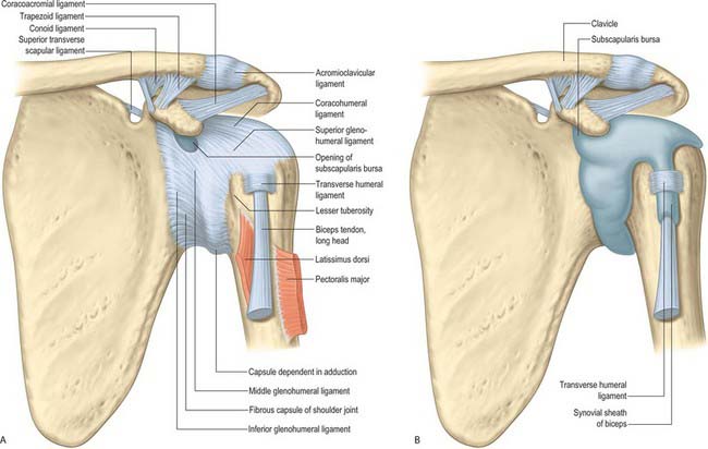
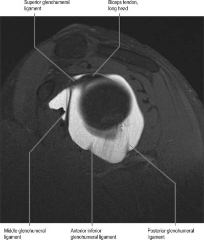
Three glenohumeral ligaments, only visible from within the joint, reinforce the capsule anteriorly and inferiorly (Fig. 46.18). They do not act as traditional ligaments (which carry a pure tensile force along their length), but become taut at varying positions of abduction and humeral rotation, acting as ‘check-reins’. Moreover, they do not have the strength characteristics of the ligaments at the knee. The superior glenohumeral ligament passes from the supraglenoid tubercle, just anterior to the origin of the long head of biceps, to the humerus near the proximal tip of the lesser tubercle on the medial ridge of the intertuberculous groove, the fovea capitis. It forms an anterior cover around the long head of biceps, and is part of the rotator interval. Together with the coracohumeral ligament it is an important stabilizer in the inferior direction, helping to keep the humeral head suspended (the coracohumeral ligament is more robust than the superior glenohumeral ligament). The middle glenohumeral ligament arises from a wide attachment below the superior glenohumeral ligament, along the anterior glenoid margin as far as the inferior third of the rim, and passes obliquely inferolaterally, enlarging as it does, to attach to the lesser tubercle deep to the tendon of subscapularis, with which it blends. The width and thickness of this ligament may be as much as 2 cm and 4 mm respectively. It provides anterior stability at 45° and 60° abduction. The thicker and longer inferior glenohumeral ligament complex is a hammock-like structure with anchor points on the anterior and posterior sides of the glenoid. It arises from the anterior, middle and posterior margins of the glenoid labrum, below the epiphysial line, and passes anteroinferiorly to the inferior and medial aspects of the neck of the humerus. The anterior, superior edge of the inferior ligament is thickened as the superior band, and the diffuse thickening of the anterior part of the capsule to which it is attached is known as the axillary pouch. The anterior band of the inferior glenohumeral ligament is thought to be the primary static anterior stabilizer of the abducted and externally rotated glenohumeral joint. (For further details consult Burkart & Debski 2002.)
The coracohumeral ligament is attached to the dorsolateral base of the coracoid process and extends as two bands, which blend with the capsule, to the greater and lesser tubercles (Fig. 46.14). Portions of the coracohumeral ligament form a tunnel for the biceps tendon on the anterior side of the joint. The rotator interval is reinforced by the coracohumeral ligament. It also blends inferiorly with the superior glenohumeral ligament.
The synovial membrane lines the capsule and covers parts of the anatomical neck. The long tendon of biceps traverses the joint in a synovial sheath which continues into the intertubercular sulcus as far as the surgical neck of the humerus (Fig. 46.14, Fig. 46.16).
Movements at the shoulder (glenohumeral) joint
In analysis of shoulder movements it is preferable to refer humeral movement to the scapula, rather than to conventional anatomical planes (Fig. 46.19). When the arm hangs at rest the glenoid fossa faces almost equally forwards and laterally, and the humeral capitular and scapular (topographical) axes correspond, although the humerus, relative to the anatomical position, is medially rotated. Flexion carries the arm anteromedially on an axis through the humeral head orthogonal to the glenoid fossa at its centre. Abduction and adduction occur in a vertical plane orthogonal to that of flexion–extension; the axis is horizontal, through the humeral head, parallel with the glenoid plane. Pure abduction raises the arm anterolaterally in the plane of the scapula. However, when referred to the trunk, flexion and extension occur in the paramedian plane, and abduction and adduction in the coronal plane. In this sense, raising the arm vertically from flexion or raising it from abduction are both accompanied by humeral rotation in opposite directions. Whether ‘scapular’ or any other plane of abduction is described, these are selections from an infinite series. In scapular abduction points on the humeral surface pursue vertical cords but in rotation they are horizontal. In ‘pure’ flexion–extension, in a plane orthogonal to the scapula the axis of movement, and the notional ‘mechanical axis’, are regarded as projected from the centre of the glenoid cavity.
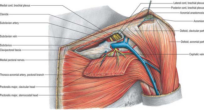
Fig. 46.19 Left deltopectoral groove showing clavipectoral fascia and thoracoacromial axis.
(From Sobotta 2006.)
The peculiar relation of the long head of biceps to the shoulder joint may serve several purposes. By its connection with both the shoulder and elbow, the muscle harmonizes their actions as an elastic ligament during all their movements. It helps to prevent the humeral head impinging on the acromion when deltoid contracts and to steady it in movements of the arm. In paralysis of supraspinatus it may also help initiate abduction of the arm, particularly when the humerus is laterally rotated.
MUSCLES
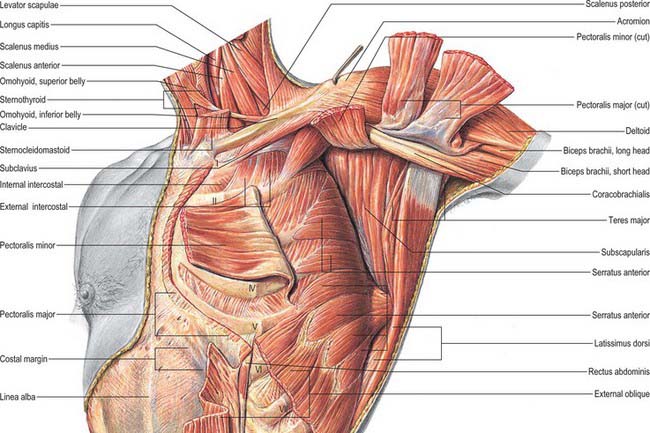
Pectoralis major
Pectoralis major (Fig. 46.19) is a thick, fan-shaped muscle. It arises from the anterior surface of the sternal half of the clavicle; half the breadth of the anterior surface of the sternum down to the level of the sixth or seventh costal cartilage; the first to the seventh costal cartilages (first and seventh often omitted); the sternal end of the sixth rib; and the aponeurosis of external oblique. The clavicular fibres are usually separated from the sternal fibres by a slight cleft. The muscle converges to a flat tendon, approximately 5 cm across, which is attached to the lateral lip of the intertubercular sulcus of the humerus. The tendon is bilaminar. The thicker anterior lamina is formed by fibres from the manubrium, which are joined superficially by clavicular fibres and deeply by fibres from the sternal margin and the second to fifth costal cartilages. Clavicular fibres may be prolonged into the deltoid tendon. The posterior lamina receives fibres from the sixth (and often seventh) costal cartilages, sixth rib, sternum, and aponeurosis of the external oblique. Costal fibres join the lamina without twisting. Fibres from the sternum and aponeurosis curve around the lower border, turning successively behind those above them, which means that this part of the muscle is twisted so that the fibres that are lowest at their medial origin are highest at their insertion on the humerus. The posterior lamina reaches higher on the humerus than the anterior, and gives off an expansion which covers the intertubercular sulcus and blends with the capsular ligament of the shoulder joint. An expansion from the deepest part of the lamina lines the intertubercular sulcus at its linear insertion; from its lower border another expansion descends into the deep fascia of the upper arm.
Poland syndrome is a rare congenital anomaly where there is hypoplasia of the thoracic chest wall muscles and ipsilateral hypoplasia of the arm and hand. It occurs in approximately 1 : 50,000 live births. The major clinical features are that the sternocostal head of pectoralis major and all of pectoralis minor are absent. In addition, there may be hypoplasia of latissimus dorsi, serratus anterior, external oblique, supraspinatus, infraspinatus, deltoid and the intercostal muscles, and hypoplasia of the hemithorax and ribs. There may be ipsilateral breast hypoplasia and absent nipple-areolar-complex (Ch. 55).
Pectoralis minor
Pectoralis minor is a thin, triangular muscle lying posterior (deep) to pectoralis major (Fig. 46.20). It arises from the upper margins and outer surfaces of the third to fifth ribs (frequently second to fourth), near their cartilages, and from the fascia over the adjoining external intercostal muscles. Its fibres ascend laterally under cover of pectoralis major, converging in a flat tendon which is attached to the medial border and upper surface of the coracoid process of the scapula. Part or all of the tendon may cross the coracoid process into the coraco-acromial ligament, or even beyond to the coracohumeral ligament, thereby gaining attachment to the humerus.
Subclavius
Subclavius is a small, triangular muscle tucked between the clavicle and the first rib (Fig. 46.20). It arises from the junction of the first rib and its costal cartilage by a thick tendon, prolonged at its inferior margin and anterior to the costoclavicular ligament. It passes upwards and laterally to a groove on the under surface of the middle third of the clavicle, where it is attached by muscular fibres.
Trapezius
Trapezius is a flat, triangular muscle which extends over the back of the neck and upper thorax (Fig. 54.15). The paired trapezius muscles form a diamond shape, from which the name is derived: the lateral angles occur at the shoulder tips, the superior angle at the occipital protuberance and superior nuchal lines, and the inferior angle at the spine of the twelfth thoracic vertebra. On either side, the muscle is attached to the medial third of the superior nuchal line, external occipital protuberance, ligamentum nuchae, and apices of the spinous processes and their supraspinous ligaments from C7 to T12. Superior fibres descend, inferior fibres ascend, and the fibres between them proceed horizontally: all converge laterally on the shoulder. The superior fibres are attached to the posterior border of the lateral third of the clavicle; the middle fibres to the medial acromial margin and superior lip of the crest of the scapular spine; and the inferior fibres pass into an aponeurosis which glides over a smooth triangular surface at the medial end of the scapular spine and is attached to a tubercle at its lateral apex. The occipital attachment is by a fibrous lamina, which is also adherent to the skin. The spinal attachment is by a broad triangular aponeurosis from the sixth cervical to the third thoracic vertebrae, and by short tendinous fibres below this.
Trapezius cooperates with other muscles in steadying the scapula, controlling it during movements of the arm, and maintaining the level and poise of the shoulder. Electromyographic activity is minimal in the unloaded arm, and heavy loads can be suspended with a small contribution from the upper part. Acting with levator scapulae, the upper fibres elevate the scapula and with it the point of the shoulder; acting with serratus anterior, trapezius rotates the scapula forward (upwards) so that the arm can be raised above the head; and acting with the rhomboids, it retracts the scapula, bracing back the shoulder. With the shoulder fixed, trapezius may bend the head and neck backwards and laterally. Trapezius, levator scapulae, rhomboids and serratus anterior combine in producing a variety of scapular rotations (p. 803).
Deltoid
Deltoid is a thick, curved triangle of muscle. It arises from the anterior border and superior surface of the lateral third of the clavicle, the lateral margin and superior surface of the acromion, and the lower edge of the crest of the scapular spine (other than its smooth medial triangular surface) (Fig. 46.19). The fibres converge inferiorly to a short, substantial tendon which is attached to the deltoid tubercle on the lateral aspect of the midshaft of the humerus. Anterior and posterior fibres converge directly to this tendon. The intermediate part is multipennate: four intramuscular septa descend from the acromion to interdigitate with three septa ascending from the deltoid tubercle. The septa are connected by short muscle fibres, which provide powerful traction. The fasciculi are large, producing a coarse longitudinal striation. The muscle surrounds the glenohumeral articulation on all sides except inferomedially, lending the shoulder its rounded profile. In contraction its borders are easily seen and felt. The tendon gives off an expansion into the brachial deep fascia which may reach the forearm.
Different parts of the muscle can act independently as well as together. Anterior fibres assist pectoralis major in drawing the arm forwards and rotating it medially. Posterior fibres act with latissimus dorsi and teres major in drawing the arm backwards and rotating it laterally. The multipennate acromial part of deltoid is a strong abductor: aided by supraspinatus it abducts the arm until the joint capsule is tense below. Movement takes place in the plane of the body of the scapula, which is the only way that scapular rotation can be fully effective in raising the arm above the head. In true abduction (Fig. 46.19), acromial fibres contract strongly, while clavicular and posterior fibres prevent departure from the plane of motion. In the early stages of abduction, traction by the deltoid is upward, but the humeral head is prevented from translating upward by the synergistic downward pull of subscapularis, infraspinatus and teres minor. Electromyography suggests that deltoid contributes little to medial or lateral rotation but confirms that it takes part in most other shoulder movements. It may also aid supraspinatus in resisting the downward drag of a loaded arm. A common action of the deltoid is arm-swinging while walking.
Levator scapulae
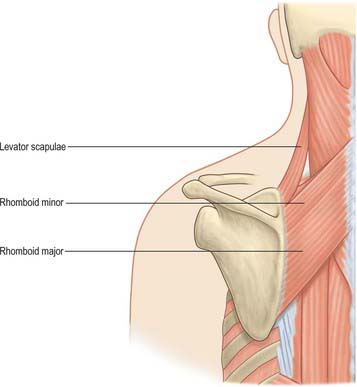
Levator scapulae is a slender muscle attached by tendinous slips to the transverse processes of the atlas and axis, and to the posterior tubercles of the transverse processes of the third and fourth cervical vertebrae (Fig. 28.6, see below Fig. 46.21). It descends diagonally to approach the medial scapular border between its superior angle and the triangular smooth surface at the medial end of the scapular spine.
Rhomboid major
Rhomboid major is a quadrilateral sheet of muscle which arises by tendinous fibres from the spines and supraspinous ligaments of the second to fifth thoracic vertebrae, and descends laterally to the medial border of the scapula between the root of the spine and the inferior angle (Fig. 46.21). Most of its fibres usually end in a tendinous band between these two points, joined to the medial border by a thin membrane. Occasionally this is incomplete, in which case some muscular fibres are attached directly into the scapula. The attachments of rhomboid major – and also those of rhomboid minor, levator scapulae and serratus anterior – can be more extensive, with ‘folds’ or extensions passing to both dorsal and costal aspects of the scapula adjacent to its medial margin.
Rhomboid minor
Rhomboid minor is a small, cylindrical muscle. It runs from the lower ligamentum nuchae and the spines of the seventh cervical and first thoracic vertebrae to the base of a smooth triangular surface at the medial end of the spine of the scapula, where dorsal and ventral layers enclose the inferior border of levator scapulae (Fig. 46.21). The dorsal layer of rhomboid minor is attached to the rim of the triangular surface, dorsolateral to and below levator scapulae. The ventral layer is strong and wide, extending 2–3 cm medial to and below levator scapulae; here the fasciae of rhomboid minor and serratus anterior are tightly fused. Rhomboid minor is usually separate from rhomboid major, but the muscles overlap and are occasionally united.
Latissimus dorsi
Latissimus dorsi is a large, flat, triangular muscle that sweeps over the lumbar region and lower thorax and converges to a narrow tendon (Fig. 46.22, Fig. 54.15). It arises by tendinous fibres from the spines of the lower six thoracic vertebrae anterior to trapezius; from the posterior layer of thoracolumbar fascia, by which it is attached to the spines and supraspinous ligaments of the lumbar and sacral vertebrae; and from the posterior part of the iliac crest. It also springs by muscular fibres from the posterior part (outer lip) of the iliac crest lateral to erector spinae, and by fleshy slips from the three or four lower ribs, interdigitating with external oblique (Fig. 61.9). From this extensive attachment, fibres pass laterally with different degrees of obliquity (the upper fibres are nearly horizontal, the middle oblique, and the lower almost vertical) to form a sheet approximately 12 or 13 mm thick that overlaps the inferior scapular angle. The muscle curves around the inferolateral border of teres major to gain its anterior surface. Here it ends as a flattened tendon, approximately 7 cm long, in front of the tendon of teres major. It is attached to the floor of the intertubercular sulcus of the humerus, with an expansion to the deep fascia. The attachment extends higher on the humerus than that of teres major. As the muscle curves round teres major, the fasciculi rotate around each other, so that fibres that originate lowest at the midline insert highest on the humerus, and fibres that originate highest at the midline insert lowest on the humerus. A bursa sometimes occurs between the muscle and the inferior scapular angle. The tendons of latissimus dorsi and teres major are united at their lower borders: they are separated by a bursa near their humeral attachments.
Latissimus dorsi is supplied by a single dominant vascular pedicle, the thoracodorsal artery, itself a continuation of the subscapular artery (p. 817). The thoracodorsal artery (and its accompanying venae comitantes) enters the muscle at a single neurovascular hilum on its costal surface, 6–12 cm from the subscapular artery and 1–4 cm medial to the lateral border of the muscle. The artery gives off one to three large branches to serratus anterior before dividing at, or even before, the hilum to supply latissimus dorsi itself. The basic pattern of branching is a bifurcation into lateral and medial branches. The larger lateral branch follows a course parallel to, and 1–4 cm from, the upper border of the muscle; the smaller branch diverges at an angle of 45° and travels medially. In a small number of cases the artery trifurcates: this usually yields a small recurrent branch which returns to supply the proximal part of the muscle, but in some cases it provides a third major branch which supplies the distal part of the muscle. Occasionally the lateral branch gives off a further collateral to serratus anterior. The major branches give off 5–9 longitudinal branches which travel distally, parallel with the muscle fibres. Musculocutaneous perforators arising from these vessels supply the overlying skin.
Serratus anterior
Serratus anterior is a large muscular sheet which curves around the thorax. It arises from an extensive costal attachment and inserts on the scapula (Fig. 46.19, Fig. 46.20). Fleshy digitations spring anteriorly from the outer surfaces and superior borders of the upper eight, nine or even ten ribs, and from fasciae which cover the intervening intercostals. They lie on a long, slightly curved, line which passes inferolaterally across the thorax. The first digitations spring from the first and second ribs and intercostal fascia, the others from a single rib, and the lower four interdigitate with the upper five slips of external oblique. The muscle follows the contour of the chest wall closely. It passes ventral to the scapula and reaches the medial border of the scapula in the following way. The first digitation encloses, and is attached to, a triangular area of both the costal and dorsal surfaces of the superior scapular angle. The next two or three digitations form a triangular sheet which is attached to the costal surface along almost its entire medial border. The lower four or five digitations converge to be attached by musculotendinous fibres to a triangular impression on the costal surface of the inferior angle: they enclose the inferior angle and are also attached to a smaller triangular part of its dorsal surface near its tip.
Supraspinatus
Supraspinatus arises from the medial two-thirds of the supraspinous fossa and from the supraspinous fascia (Fig. 46.22). The fibres converge, under the acromion, into a tendon which crosses above the shoulder joint and is attached to the highest facet of the greater tubercle of the humerus. The tendon blends into the articular capsule and may give a slip to the tendon of pectoralis major. Fibrocartilage has been described at the tendinous insertion, as in other tendons attached to epiphysial bone.
Infraspinatus
Infraspinatus is a thick triangular muscle which occupies most of the infraspinous fossa (Fig. 46.22). It arises by muscular fibres from the medial two-thirds of the fossa, by tendinous fibres from ridges on its surface and from the deep surface of the infraspinous fascia, which separates it from teres major and minor. Its fibres converge to a tendon which glides under the lateral border of the spine of the scapula, and then passes across the posterior aspect of the capsule of the shoulder joint to be attached to the middle facet on the greater tubercle of the humerus. The tendon is sometimes separated from the capsule by a bursa, which may communicate with the joint cavity. Infraspinatus is sometimes fused with teres minor.
Subscapularis
Subscapularis is a bulky, triangular muscle which fills the subscapular fossa (Fig. 46.20). In its medial two-thirds, the fibres are attached to the periosteum of the costal surface of the scapula. Other fibres arise from tendinous intramuscular septa, which are attached to ridges on the bone, and from the aponeurosis which covers the muscle and separates it from teres major and the long head of triceps. The fibres converge laterally into a broad tendon which is attached to the lesser tubercle of the humerus and the front of the articular capsule. The tendon is separated from the neck of the scapula by the large subscapular bursa, which communicates with the shoulder joint.
Teres major
Teres major is a thick, flat muscle which arises from the oval area on the dorsal surface of the inferior scapular angle, and from the fibrous septa interposed between the muscle and teres minor and infraspinatus (Fig. 46.22). Its fibres ascend laterally and end in a flat tendon, approximately 5 cm long, which is attached to the medial lip of the intertubercular sulcus of the humerus. The tendon lies behind that of latissimus dorsi, from which it is separated by a bursa; the tendons are united along their lower borders for a short distance.
Teres minor
Teres minor is a narrow elongated muscle which arises from the upper two-thirds of a flattened strip on the dorsal surface of the scapula adjoining its lateral border, and from two aponeurotic laminae which separate it from infraspinatus and teres major (Fig. 46.22). It runs upwards and laterally. The upper fibres end in a tendon attached to the lowest facet on the greater tubercle of the humerus. The lower fibres are attached directly into the humerus distal to this facet and above the origin of the lateral head of triceps. The tendon passes across, and blends with, the lower posterior surface of the capsule of the shoulder joint.
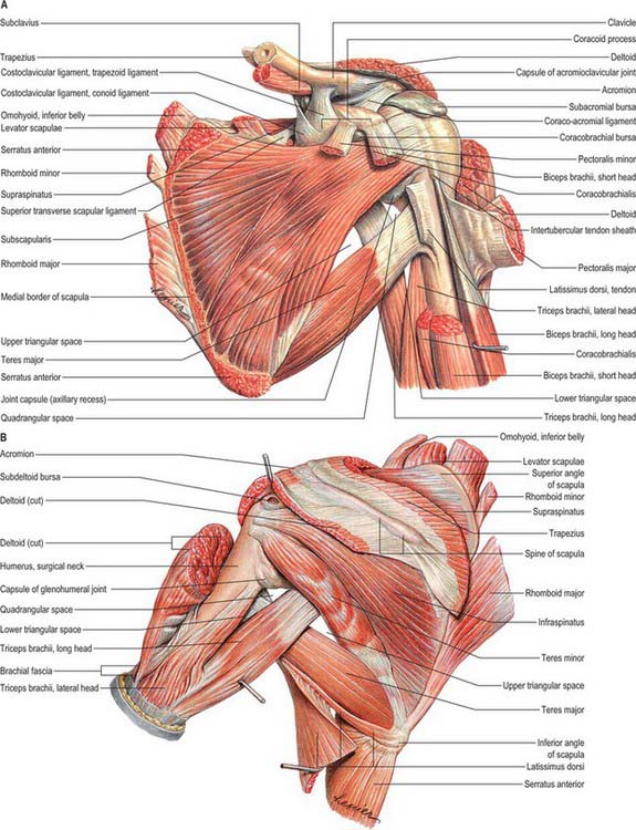
The quadrangular and triangular spaces
Anteriorly, the quadrangular space is bounded by subscapularis, the capsule of the shoulder joint and teres minor above, teres major below, the long head of triceps medially, and the surgical neck of the humerus laterally. Posteriorly, the quadrangular space is bounded above by teres minor. The axillary nerve and the posterior circumflex artery and vein pass through the space (Fig. 46.22).
There are two triangular spaces (Fig. 46.22). The upper triangular space is bounded above by subscapularis anteriorly, teres minor posteriorly, teres major below, and the long head of triceps laterally. The circumflex scapular artery passes through this space. The lower triangular space (triangular interval) is bounded above by subscapularis anteriorly and teres major posteriorly; the long head of triceps medially and the humerus laterally. The radial nerve and the profunda brachii vessels pass through this space.
AXILLA
BOUNDARIES
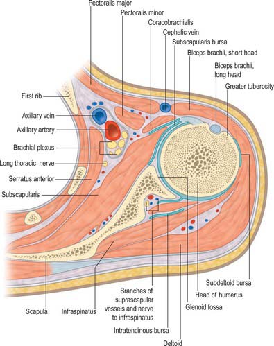
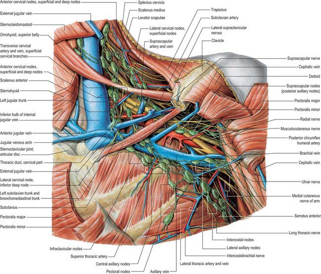
The axilla is a pyramidal region between the upper thoracic wall and the arm (Fig. 46.23, Fig. 46.24). Its blunt apex continues into the root of the neck (cervico-axillary canal) between the external border of the first rib, superior border of the scapula, posterior surface of the clavicle and the medial aspect of the coracoid process. Its base, which is virtual, can be imagined as facing downwards: it is broad at the chest and narrow at the arm, and corresponds to the skin and a thick layer of axillary fascia between the inferior borders of pectoralis major anteriorly and latissimus dorsi posteriorly. It is convex upwards, conforming to the concavity of the armpit.
VASCULAR SUPPLY AND LYMPHATIC DRAINAGE
ARTERIES
Suprascapular artery
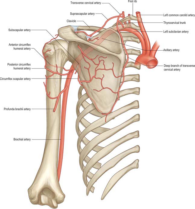
The suprascapular artery usually arises from the thyrocervical trunk of the subclavian artery, although it may arise from the third part of the subclavian artery (Fig. 28.7). It first descends laterally across scalenus anterior and the phrenic nerve, posterior to the internal jugular vein and sternocleidomastoid, then crosses anterior to the subclavian artery and brachial plexus, posterior and parallel with the clavicle and subclavius and the inferior belly of omohyoid, to reach the superior border of the scapula. Here it passes above (sometimes under) the superior transverse ligament, separating it from the suprascapular nerve, and enters the supraspinous fossa (Fig. 46.25), where it lies on the bone, and supplies supraspinatus. It descends behind the scapular neck, and passes through the spinoglenoid notch deep to the inferior transverse ligament to gain the deep surface of infraspinatus, where it anastomoses with the circumflex scapular and deep branch of the transverse cervical artery. Its muscular branches supply sternocleidomastoid, subclavius and infraspinatus. It also gives off a suprasternal branch which crosses the sternal end of the clavicle to supply the skin of the upper thorax, and an acromial branch which pierces trapezius to supply the skin over the shoulder. This last branch anastomoses with the thoraco-acromial and posterior circumflex humeral arteries.
Dorsal scapular artery
In the majority of cases, the dorsal scapular artery arises from the third, or less often the second, part of the subclavian artery. It passes laterally through the trunks of the brachial plexus in front of scalenus medius and then deep to levator scapulae to reach the superior scapular angle. Here it descends with the dorsal scapular nerve under the rhomboids along the medial border of the scapula to the inferior angle (Fig. 46.25). It supplies the rhomboids, latissimus dorsi and the inferior portion of trapezius. It supplies the skin over the inferomedial aspect of trapezius via musculocutaneous perforators. It anastomoses with the suprascapular and subscapular and with posterior branches of some posterior intercostal arteries. It sends a small branch to scalenus anterior: sometimes this arises directly from the subclavian artery.
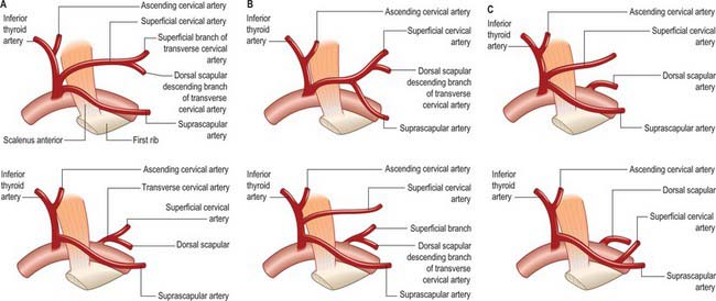
About a third of the superficial cervical and dorsal scapular arteries arise in common from the thyrocervical trunk as a transverse cervical artery, together with a superficial and a deep branch (superficial cervical and dorsal scapular arteries respectively). In this case the dorsal scapular artery arises near the superior border of the scapula: it passes laterally, anterior to the brachial plexus and then posterior to levator scapulae (Fig. 46.26).
Axillary artery
Relations of the third part
Pectoralis major, and, distal to the muscle, skin and fasciae, are anterior. The lower part of subscapularis and the tendons of latissimus dorsi and teres major are posterior. Coracobrachialis is lateral and the axillary vein is medial. Branches of the brachial plexus are arranged as follows: laterally, the lateral root and then trunk of the median nerve and, for a short distance, the musculocutaneous nerve; medially, the medial cutaneous nerve of the forearm between the axillary artery and vein anteriorly, and the ulnar nerve between these vessels posteriorly; anteriorly, the medial root of the median nerve; posteriorly, the radial and axillary nerves, the latter only to the distal border of subscapularis.
Branches
Superior thoracic artery
The superior thoracic artery is a small vessel which arises from the first part of the axillary artery near the lower border of subclavius: it sometimes arises from the thoraco-acromial artery (Fig. 46.24). It runs anteromedially above the medial border of pectoralis minor, then passes between it and pectoralis major to gain the thoracic wall. It supplies these muscles and the thoracic wall, and anastomoses with the internal thoracic and upper intercostal arteries.
Thoraco-acromial (acromio-thoracic) artery
The thoraco-acromial artery is a short branch which arises from the second part of the axillary artery (Fig. 28.7, Fig. 46.19). It is at first overlapped by pectoralis minor; skirting its medial border, next pierces the clavipectoral fascia and divides into pectoral, acromial, clavicular and deltoid branches, which supply pectoralis major and minor, an area of skin over the clavipectoral fascia and the anterior portion of deltoid.
Subscapular artery
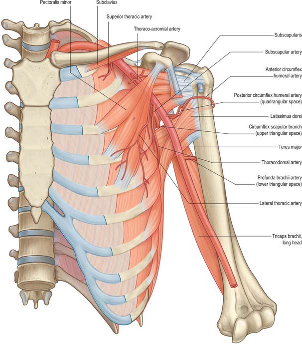
The subscapular artery is the largest branch of the axillary artery (Fig. 46.24, Fig. 46.25). It usually arises from the third part of the axillary artery at the distal (inferior) border of subscapularis, which it follows to the inferior scapular angle, where it anastomoses with the lateral thoracic and intercostal arteries and the deep branch of the transverse cervical artery. It supplies adjacent muscles and the thoracic wall. It is accompanied distally by the nerve to latissimus dorsi. Approximately 4 cm from its origin the subscapular artery divides into the circumflex scapular and thoracodorsal arteries.
Posterior circumflex humeral artery
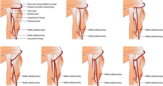
The posterior circumflex humeral artery is larger than the anterior (Fig. 46.25, Fig. 46.27). It branches from the third part of the axillary artery at the distal border of subscapularis and runs backwards with the axillary nerve through a quadrangular space which is bounded by subscapularis, the capsule of the shoulder joint and teres minor above, teres major below, the long head of triceps medially, and the surgical neck of the humerus laterally. It curves round the humeral neck and supplies the shoulder joint, deltoid, teres major and minor, and long and lateral heads of triceps. It gives off a descending branch which anastomoses with the deltoid branch of the profunda brachii artery and with the anterior circumflex humeral and acromial branches of the suprascapular and thoraco-acromial arteries.
VEINS
Axillary vein
The axillary vein is the continuation of the basilic vein. It begins at the lower border of teres major, and ascends to the outer border of the first rib, where it becomes the subclavian vein. It is joined by the brachial vein near subscapularis, and by the cephalic vein near its costal end; other tributaries follow the axillary arterial branches. It lies medial to the axillary artery, which it partly overlaps. The medial pectoral nerve, medial cord of the brachial plexus, ulnar nerve and medial cutaneous nerve of the forearm lie between the artery and the vein. The medial cutaneous nerve of the arm is medial to the vein; the lateral group of axillary lymph nodes is posteromedial. There are a pair of valves near its distal end, and valves also occur near the ends of the cephalic and subscapular veins.
AXILLARY LYMPH NODES
There are between 20 to 30 axillary nodes, which may be divided into five not wholly distinct groups, namely, lateral, anterior (pectoral), posterior (subscapular), central and apical (Fig. 54.21, Fig. 45.6, Fig. 46.28). Four of the groups are intermediary, and only the apical group is terminal. Collectively they drain the entire upper limb, breast and trunk above the umbilicus.
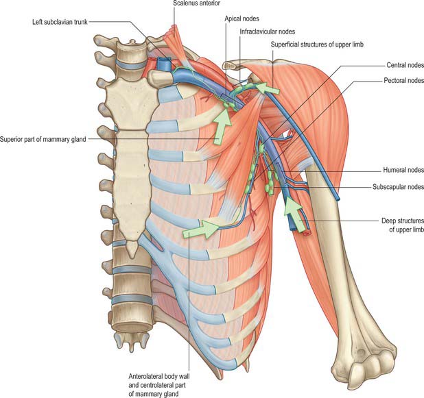
The lateral group of four to six nodes is posteromedial to the axillary vein, its afferents drain the whole limb except the vessels accompanying the cephalic vein (Fig. 54.21, Fig. 45.6, Fig. 46.28). Efferent vessels pass partly to the central and apical axillary groups, and partly to the inferior deep cervical nodes. The anterior group of four or five nodes spreads along the inferior border of pectoralis minor near the lateral thoracic vessels. Their afferents drain the skin and muscles of the supraumbilical anterolateral body wall and breast, and efferents pass partly to the central and partly to the apical axillary nodes. The posterior group of six or seven nodes lie on the inferior margin of the posterior axillary wall, along the subscapular vessels. Their afferents drain the skin and superficial muscles of the inferior posterior region of the neck and the dorsal aspect of the trunk down to the iliac crest, and efferents pass to the apical and central axillary nodes. A central group of three or four large nodes embedded in axillary fat receives afferents from all preceding groups, and their efferents drain to the apical nodes. An apical group of six to twelve nodes is partly posterior to the superior part of pectoralis minor and partly above its superior border, extending to the apex of the axilla medial to the axillary vein. The only direct territorial afferents are those which accompany the cephalic vein or some which drain the upper peripheral region of the breast: the group drains all the other axillary nodes. Their efferents unite as the subclavian trunk and drain directly to the jugulosubclavian venous junction, or the subclavian vein, or jugular lymphatic trunk or (occasionally) to a right lymphatic duct; the left trunk usually ends in the thoracic duct. A few efferents from apical nodes usually reach the inferior deep cervical nodes.
INNERVATION
BRACHIAL PLEXUS
For an overview of the brachial plexus see page 779 and Fig. 45.7.
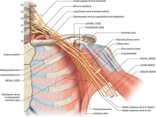
In the axilla, the lateral and posterior cords of the brachial plexus are lateral to the first part of the axillary artery, and the medial cord is behind the artery. The cords surround the second part of the artery, their names indicating their relationship. In the lower axilla the cords divide into nerves that supply the upper limb (Fig. 46.29). Other than the medial root of the median nerve, these nerves are related to the third part of the artery in the same way as their cords are related to the second part, i.e. branches of the lateral cord are lateral to the artery, branches of the medial cord are medial, and branches of the posterior cord are posterior.
Branches of the brachial plexus may be described as supraclavicular and infraclavicular.
Supraclavicular branches
Supraclavicular branches arise from roots or from trunks as follows:
| From roots | 1. Nerves to scaleni and longus colli | C5, 6, 7, 8 |
| 2. Branch to phrenic nerve | C5 | |
| 3. Dorsal scapular nerve | C5 | |
| 4. Long thoracic nerve | C5, 6 (7) | |
| From trunks | 1. Nerve to subclavius | C5, 6 |
| 2. Suprascapular nerve | C5, 6 |
Long thoracic nerve
The long thoracic nerve is usually formed by roots from the fifth to the seventh cervical rami, although the last ramus may be absent (Fig. 46.30). The upper two roots pierce scalenus medius obliquely, uniting in or lateral to it. The nerve descends dorsal to the brachial plexus and the first part of the axillary artery and crosses the superior border of serratus anterior to reach its lateral surface. It may be joined by the root from C7, which emerges between scalenus anterior and scalenus medius, and descends on the lateral surface of medius. The nerve continues downwards to the lower border of serratus anterior, and supplies branches to each of its digitations.
Suprascapular nerve
The suprascapular nerve is a large branch of the superior trunk (Fig. 28.17). It runs laterally, deep to trapezius and omohyoid and enters the supraspinous fossa through the suprascapular notch inferior to the superior transverse scapular ligament. It runs deep to supraspinatus, supplies it, and curves round the lateral border of the spine of the scapula with the suprascapular artery to reach the infraspinous fossa, where it gives two branches to infraspinatus and articular rami to the shoulder and acromioclavicular joints. The suprascapular nerve rarely has a cutaneous branch. When present, it pierces deltoid close to the tip of the acromion and supplies the skin of the proximal third of the arm within the territory of the axillary nerve.
Infraclavicular branches
| Lateral cord | Lateral pectoral | C5, 6, 7 |
| Musculocutaneous | C5, 6 7 | |
| Lateral root of median | C(5), 6, 7 | |
| Medial cord | Medial pectoral | C8, T1 |
| Medial cutaneous of forearm | C8, T1 | |
| Medial cutaneous of arm | C8, T1 | |
| Ulnar | C(7), 8, T1 | |
| Medial root of median | C8, T1 | |
| Posterior cord | Upper subscapular | C5, 6 |
| Thoracodorsal | C6, 7,8 | |
| Lower subscapular | C5, 6 | |
| Axillary | C5, 6 | |
| Radial | C5, 6, 7, 8, (T1) |
Lateral pectoral nerve
The lateral pectoral nerve is larger than the medial, and may arise from the anterior divisions of the upper and middle trunks, or by a single root from the lateral cord. Its axons are from the fifth to seventh cervical rami. It crosses anterior to the axillary artery and vein, pierces the clavipectoral fascia and supplies the deep surface of pectoralis major. It sends a branch to the medial pectoral nerve, forming a loop in front of the first part of the axillary artery (Fig. 46.30), to supply some fibres to pectoralis minor.
Axillary nerve
The axillary nerve arises from the posterior cord (C5, 6) (Fig. 46.22). It is at first lateral to the radial nerve, posterior to the axillary artery and anterior to subscapularis. At the lower border of subscapularis it curves back inferior to the humeroscapular articular capsule and, with the posterior circumflex humeral vessels, traverses a quadrangular space bounded above by subscapularis (anterior) and teres minor (posterior), below by teres major, medially by the long head of triceps, and laterally by the surgical neck of the humerus. It divides in the space into anterior and posterior branches. The anterior branch curves round the neck of the humerus with the posterior circumflex humeral vessels, deep to deltoid. It reaches the anterior border of the muscle and supplies it, and gives off a few small cutaneous branches which pierce deltoid and ramify in the skin over its lower part. The posterior branch courses medially and posteriorly along the attachment of the lateral head of triceps, inferior to the glenoid rim. It usually lies medial to the anterior branch in the quadrangular space. It gives off the nerve to teres minor and the upper lateral cutaneous nerve of the arm at the lateral edge of the origin of the long head of triceps. The nerve to teres minor enters the muscle on its inferior surface. The posterior branch frequently supplies the posterior aspect of deltoid, usually via a separate branch from the main stem, occasionally from the superior lateral cutaneous nerve of the arm. However, the posterior part of deltoid has a more consistent supply from the anterior branch of the axillary nerve, which should be remembered when performing a posterior deltoid-splitting approach to the shoulder. The upper lateral cutaneous nerve of the arm pierces the deep fascia at the medial border of the posterior aspect of deltoid and supplies the skin over the lower part of deltoid and the upper part of the long head of triceps. The posterior branch is intimately related to the inferior aspects of the glenoid and shoulder joint capsule, which may place it at particular risk during capsular plication or thermal shrinkage procedures (Ball et al 2003). There is often an enlargement or pseudoganglion on the branch to teres minor. The axillary trunk supplies a branch to the shoulder joint below subscapularis.
Musculocutaneous nerve
The musculocutaneous nerve arises from the lateral cord (C5–7), opposite the lower border of pectoralis minor (Fig. 46.30). It pierces coracobrachialis and descends laterally between biceps and brachialis to the lateral side of the arm. Just below the elbow it pierces the deep fascia lateral to the tendon of biceps, and continues as the lateral cutaneous nerve of the forearm. A line drawn from the lateral side of the third part of the axillary artery across coracobrachialis and biceps to the lateral side of the biceps tendon is a surface projection for the nerve (but this varies according to its point of entry into coracobrachialis). It supplies coracobrachialis, both heads of biceps and most of brachialis. The branch to coracobrachialis is given off before the musculocutaneous nerve enters the muscle: its fibres are from the seventh cervical ramus and may branch directly from the lateral cord. Branches to biceps and brachialis leave after the musculocutaneous has pierced coracobrachialis: the branch to brachialis also supplies the elbow joint. The musculocutaneous nerve supplies a small branch to the humerus, which enters the shaft with the nutrient artery.
Medial cutaneous nerve of forearm
The medial cutaneous nerve of the forearm arises from the medial cord (C8, T1). It is described further on pages 830 and 856.
Median nerve
The median nerve has two roots from the lateral (C5, 6, 7) and medial (C8, T1) cords, which embrace the third part of the axillary artery, and unite anterior or lateral to it (Fig. 46.30). Some fibres from C7 often leave the lateral root in the lower part of the axilla and pass distomedially posterior to the medial root, and usually anterior to the axillary artery, to join the ulnar nerve. They may branch from the seventh cervical ventral ramus. Clinically they are believed to be mainly motor and to supply flexor carpi ulnaris. If the lateral root is small, the musculocutaneous nerve (C5, 6, 7) connects with the median nerve in the arm.
Ulnar nerve
The ulnar nerve arises from the medial cord (C8, T1) but often receives fibres from the ventral ramus of C7 (Fig. 46.30). It runs distally through the axilla medial to the axillary artery, between it and the vein. It is described further on page 827.
Radial nerve
The radial nerve is the largest branch of the brachial plexus. It arises from the posterior cord (C5, 6, 7, 8, (T1)) (Fig. 46.22), and descends behind the third part of the axillary artery and the upper part of the brachial artery, anterior to subscapularis and the tendons of latissimus dorsi and teres major. With the profunda brachii artery it inclines dorsally, and passes through the triangular space below the lower border of teres major, between the long head of triceps and the humerus. It is described further on page 827.
Ball CM, Steger T, Galatz LM, Yamaguchi K. The posterior branch of the axillary nerve: an anatomic study. J Bone Joint Surg. 2003;85:1497-1501.
Burkart AC, Debski RE. Anatomy and function of the glenohumeral ligaments in anterior shoulder instability. Clin Orthopaed Related Res. 2002;400:32-39.
Michael Salmon: Anatomic Studies. In: Taylor GI, Razaboni RM. Book 1, Arteries of the Muscles of the Extremities and the Trunk, Book 2, Arterial anastomotic pathways of the extremities. St Louis: Quality Medical Publishing, 1994.