Pattern Dystrophy
Clinical Features:
There are dark, yellow, and/or orange pigment disturbances at the level of the RPE, leading to characteristic patterns of deposition in the central macula, which are often vitelliform-like (Fig. 12.3.1). There is a lifetime risk up to 18% of developing secondary choroidal neovascularization. The clinical features are usually symmetric, but there can be heterogeneity between eyes (Fig. 12.3.2).
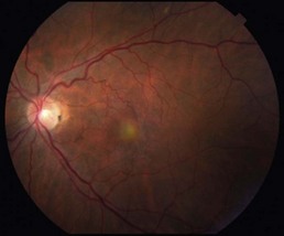
Figure 12.3.1 Color fundus photograph shows a yellowish, circular vitelliform-like lesion within the central macula.
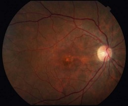
Figure 12.3.2 Color fundus photograph of fellow eye from Figure 12.3.1, shows numerous clumps of pigment within the central macula. This likely represents a collapsed vitelliform lesion.
OCT Features:
A disturbance at the level of the RPE is the rule. In the setting of a vitelliform-like lesion, there is moderately reflective material underneath or within the RPE layer (Fig. 12.3.3). Below this are highly reflective, drusen-like deposits. In the absence of a vitelliform-like lesion, there are typically highly reflective, drusen-like deposits within the RPE layer (Fig. 12.3.4). There can be a hyporeflective, empty space overlying the pigmentary disturbance. The significance of this fluid-like compartment is not clearly understood, but it usually does not represent the presence of choroidal neovascularization and would not be expected to be VEGF-responsive.
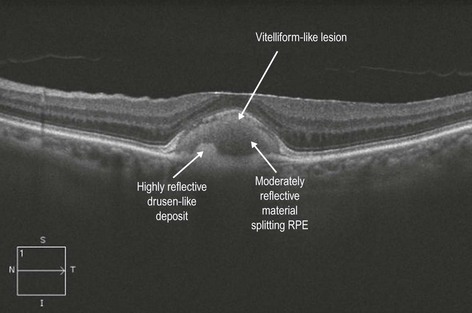
Figure 12.3.3 OCT (corresponding to Figure 12.3.1) shows moderately reflective material that appears to split the RPE layer. There are also highly reflective, drusen-like deposits underlying this area.
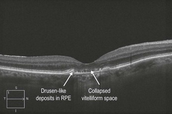
Figure 12.3.4 OCT (corresponding to Figure 12.3.2) shows highly reflective, drusen-like deposits corresponding to the pigment disturbances seen in the color photograph.
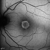
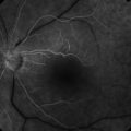
Ancillary Testing:
Fluorescein angiography can be helpful in ruling out secondary choroidal neovascularization, particularly when OCT reveals the presence of a fluid-like subretinal compartment. Fundus autofluorescence often has a characteristic appearance that can aid in the diagnosis (Fig. 12.3.5).