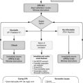
Chapter 29. Patient immobilisation and extrication
It is crucial that decisions about stabilisation, extrication and subsequent evacuation are taken early on in the rescue. The techniques used for stabilisation must not be viewed in isolation, but should be part of the total rescue activity and should complement the other treatments used.
Principles of immobilisation
In the prehospital setting, the principles of skeletal management are to:
• Prevent further injury
• Ensure neurovascular supply
• Make the patient comfortable.
The overriding importance of managing the airway with cervical spine protection, breathing and circulation (ABC) is fundamental to the treatment of any injury.
With the exception of cervical spine care, fracture management and extrication follow the primary survey unless a ‘snatch rescue’ is necessary.
The principles of definitive fracture management are:
• Reduction
• Immobilisation
• Preservation of function.
Every piece of equipment will cover or hide the patient to a greater or lesser extent, it is essential that any immediate local treatment and observations are carried out before the immobilisation device is applied.
Wounds should wherever possible be photographed and appropriately dressed before immobilisation.
The benefits of immobilisation are:
• Pain relief
• Reduction of blood loss
• Prevention of neurovascular damage
• Prevention of fat embolism.
Remembering the ABC principles, splinting or extrication devices should never produce any airway, breathing or circulation compromise.
The patient should always feel more comfortable after the splint or device has been applied, so that handling becomes easier.
Always check the pulses distal to an injury before starting treatment
Forms of splintage
Box splints
Box splints are simple in design and are useful for some arm, lower leg and ankle injuries and are carried by every front-line ambulance.
The splint forms an oblong box, open along one side with the other three sides able to be folded in such a way as to form a gutter. There may be a foot support at one end. Box splints are available in adult and child sizes.
Application
• Expose the injured leg
• Remove footwear (occasionally, footwear may provide support and should be left in place)
• Apply dressings to any wounds
• Straighten the ankle and check the peripheral pulses
• Raise the leg and pass the splint passed underneath it
• Fold the two sides of the splint so they fit closely against the leg, place the foot support
• Secure with the Velcro straps
• If any strap passes near to an injury, care should be taken that it does not cause pain; if it does, it should be left loose
• Once the splint is applied, the patient should be rechecked – specifically, the pulses in the limb and the distal sensation must be noted and recorded
• Mark the position of a palpable dorsalis pedis pulse with pen once a splint has been applied to the leg.
Traction splints
The primary function of a traction splint is to immobilise the fracture (of a lower limb) in a reduced position
• Following a fracture of the shaft of the femur the muscles of the thigh will shorten the leg, causing the bone ends to override. This increases the radius of the thigh so that it becomes more spherical: this shape has a larger internal volume than a cylinder and so presents a larger space into which blood can fill
• Application of traction will restore the cylindrical shape of the thigh, reducing its volume and reducing the overall blood loss
• Three types of traction splint are found in prehospital care: the Hare® or Trac-3® splint, the Sager® splint and the Donway® splint
• There may be other more life-threatening injuries which must take priority. Patients are far less likely to die from a fracture of the lower limb than they are from a blocked airway
• All compound fractures will need to be explored and cleaned. Therefore the receiving hospital must know that a fracture was compound and if possible a photograph should be taken.
Indications for traction splintage
• Closed (simple) fractures of the femoral shaft
• Closed (simple) fractures of the proximal two-thirds of the tibia and fibula
• Compound fractures of the femur, and the proximal two-thirds of the tibia and fibula.
Contraindications to traction splintage
• Fractures around the knee
• Dislocation of the hip
• Fracture dislocation of the knee
• Ankle injuries
• Simple undisplaced fracture of the lower third of the tibia and fibula (better immobilised with a box splint)
• Fractures of the pelvis.
Complications of traction splintage
• Damage to the neurovascular supply to the leg. This can be prevented by careful examination of the distal limb function
• Absence or change in distal function must be reported to the Emergency Department. If it is found that the distal pulses diminish or are absent after traction has been applied then traction must be gently reduced until the pulse returns
• The pulse oximeter can be used to detect alterations in the blood flow if the probe is placed on one of the toes of the fractured leg.
Hare® or Trac-3® traction splint
This splint can be used with traction to maintain a reduced fracture of the lower limb and can also be used without traction simply for support.
Application
• The splint requires two people to apply it correctly and analgesia should be given as required before manipulating the fracture
• The splint should be set-up as follows:
• The fracture site is exposed (clothes should be cut if necessary). Motorcycle leathers should not be removed as these can be dramatically effective in the control of lower limb and pelvic fracture bleeding
• The limb should be examined thoroughly and the footwear removed
• The pulses distal to the fracture together with the colour and warmth of the limb and sensation and motor function distal to the fracture should also be assessed (the neurovascular examination)
• Wounds are dressed if required
• The splint is prepared
• Select the appropriate ankle hitch
• The splint is placed by the good leg, measured for length and adjusted accordingly, then laid by the injured leg
• All the straps are checked; these should be open and placed at the correct intervals down the splint
• Some of the traction strap should be unwound
• The foot is straightened and the ankle hitch placed well under the ankle
• The side straps are then tightly folded over the ankle (not around the foot) and the rings brought together below the foot
• Finally the strap at the bottom of the foot is firmly grasped: traction must be applied along the longitudinal axis of the femur, not over the dorsum of the foot, which can cause permanent damage to the limb
• Manual traction is started with one hand while the other hand supports the leg
• The splint is then put in the correct position. The best method is to roll the patient away from the splint while a colleague slides the splint under the leg
• The top padded ring must fit under the ischial tuberosity. The patient is then rolled back onto the splint. If the position is still not correct then the patient can be moved down slightly so that he is sitting on the padded ring. Manual traction MUST be maintained THROUGHOUT this procedure
• The top strap is done up and padding applied if required. The external genitalia should be avoided in males. If correctly positioned, this strap will lie parallel to the crease of the groin
• The traction hook is then put through the ‘D’ rings and traction taken up, ensuring that manual traction is not released before the splint’s mechanical traction is tightened
• Traction is applied until the limb is comfortable (to a maximum of 7 kg in adults)
• The neurovascular examination is repeated and the oximeter reading rechecked
• The leg is elevated by raising the foot stand
• The Velcro straps are positioned and tightened to support the site of the fracture
• The leg is covered to keep it warm.
En route to hospital
The neurovascular examination should be repeated every 5–10 minutes. The straps should be checked and loosened if required – the leg may swell.
The tension of traction should be checked; as a result of reduced spasm in the muscles, tension can be lost.
To release traction
The two splints (Hare® and Trac-3®) have slightly different release mechanisms
Manual traction is taken up and then the mechanical traction is released after all the supporting Velcro straps have been removed
The Hare® splint has a pull ring which releases the traction suddenly, whereas the Trac-3® has a knob which has to be unwound to release the traction (which is less likely to be accidentally released).
Sager® traction splint
The Sager® traction splint weighs <2 kg and can be used to treat single or bilateral fractures of the lower limb, especially of the femur.
Application
• The shoe and sock are removed and the leg exposed as necessary
• The distal pulses and sensation in the injured leg are assessed
• The cushioned end of the splint is applied between the patient’s legs, against the perineum and symphysis pubis (avoiding the external genitalia in males)
• The bridle ‘S’ strap is applied around the top of the thigh
• The splint is extended so that the ankle hitch lies between the patient’s heels or at the level of the normal heel if the fractured leg has been shortened
• The ankle harness is applied beneath the heel and wrapped around just above the malleoli, adjusting the cushions on the strap to fit the size of the leg
• Traction is applied (recommended at 10% of the body weight) until the patient is comfortable
• The leg cravats are applied
• The bridle around the thigh is tightened if necessary
• The cravats are secured
• The foot-binding strap is placed around the feet and ankles in a figure-of-eight
• The foot pulses must be checked following application.
To release traction
Manual traction is taken up. The cravats and ankle hitch are removed. Along the shaft of the splint there is a small sprung piece of metal, which should be lifted to release the tension.
Donway® splint
The Donway® splint employs a different method to achieve traction.
• The fractured leg is cradled by the splint with the foot firmly fixed to the ankle support
• The top strapping is put around the thigh and then, using the pump provided, the two halves of the splint (lower and upper) are pushed apart by increased pressure (like the slide on a trombone)
• Once the patient is comfortable the securing screws are tightened; the pressure in the splint is then released through a valve
• The leg straps are applied and (as with other forms of traction splint) the pulses and sensation in the limb must be checked.
Care must be taken to ensure the ankle hitch is applied in a manner that avoids traction over the dorsum of the foot.
Cervical spine immobilisation
Manual methods
From behind the patient:
• The rescuer’s palms are placed behind the patient’s ears
• The little fingers should lie just under the angle of the jaw and the thumbs should be extended upwards behind the posterior aspect of the skull
• The rescuer’s hands should be adjusted so that the patient’s ears lie between the fingers. It is important not to cover the ears: the last thing an anxious patient needs is to be prevented from hearing
• If the head is not in a neutral position, it should be moved slowly and gently into a neutral position. If resistance is felt during this procedure the patient should be managed in the position in which he has been found
• Traction is not applied
• The rescuer should move into a comfortable braced position in order to support their own arms to prevent them from becoming tired.
From the side:
• One hand should be placed behind the patient’s head so the occiput lies in its palm
• The other hand should support the jaw between the thumb and second finger
The two hands now hold the neck in a similar way to a cervical collar
• The head can be moved into a neutral position. The anterior arm should be braced against the patient’s sternum.
From the front:
• The rescuer’s hands are placed over the patient’s cheeks so that the fingers pass around the neck and the extended thumbs lie just in front of the ears over the temporomandibular joint
• The head may be moved into a neutral position. The anterior arm should be braced against the patient’s sternum.
Cervical collars
A number of different types of cervical collars are available. Some are ‘one piece’ and require a range of sizes to be carried (e.g. the Stiffneck® collar); others come in one piece but are adjustable. The collars have to be sized according to the manufacturer’s instructions and then applied correctly while maintaining manual immobilisation.
Collars do not completely immobilise the cervical spine and it is essential to continue manual immobilisation until this is replaced by a short spine board or long spine board and head immobiliser. It is of little use merely to hold on to the collar: the hands have to support the head and should be placed above the collar. A correctly sized collar will reduce flexion and extension and to lesser extent sideways movement, but will not stop rotation – it is rotation that the rescuer’s hands or headblock and tape will prevent.
Access to the airway and trachea is available at all times through the gap in front of the collar. The application of a cervical collar, even when correctly sized, will cause a rise in intracranial pressure (through venous compression). It is, therefore, acceptable to release the collar in these circumstances once head blocks and tape have been correctly applied.
Patients who are unconscious should have their cervical spine protected and be fully immobilised. However, patients who are conscious should be carefully assessed to include:
• Consideration of the mechanism of injury
• The presence of drugs or alcohol
• A long bone injury or other distracting injury
• The presence of midline cervical spine tenderness.
The presence of any of these features following an accident with possible cervical spine injury should mandate full immobilisation. Similarly, patients who complain of pain and are reluctant to move their neck of their own volition should receive full immobilisation.
Correct scene and patient assessment will ensure the correct management of the cervical spine.
Log roll technique
Log rolling is a method of turning patients either to inspect their backs or to help put them on to a long spinal board. The object is to keep the whole spine in alignment.
• To log roll a patient, there should be a minimum of four people
• The patient should lie with his arms by his sides and the palms placed against the legs. Alternatively the patient can place his arms in a crossed position on his chest
• The cervical spine is stabilised and the patient moved with the neck, shoulders and pelvis kept in the same plane
• One person takes the head and this person controls the manoeuvre
• The next person grips the patient’s shoulder on the opposite side and also the further arm
• The third person grips the pelvis
• The fourth person controls the legs
• The person at the head calls the instructions and the whole body is rolled over, keeping the spine from twisting
• The roll should be only as far as is needed to inspect the back or insert a long board underneath the patient
• A fifth person, if available, should examine the patient’s back and perform any necessary treatment (e.g. dressing a bleeding wound).
The patient is then rolled back into the supine position, again controlled by the person controlling the head and neck
The person at the head should inform his colleagues what command will be given before the manoeuvre begins: ‘I will say one, two, three, move. Everybody ready? Good. One, two …’.
Scoop stretcher
The scoop stretcher provides a means of lifting a patient onto a trolley or ambulance cot with minimal movement. The scoop stretcher can be split in half longitudinally and may have a head cushion. The bottom half can be extended to fit the patient
• The scoop should be laid beside the patient and extended to the required length
• The patient should be told what is about to happen
• The halves are slid underneath the patient’s body from the sides, taking care not to pinch the body as the halves are brought together
• The patient may have to be rolled slightly to allow each half stretcher to be slid underneath
• Once the stretcher is in place and the halves are locked together, the head cushion is secured and the patient is lifted onto the trolley or cot
• The distance that the stretcher has to be carried should be kept to a minimum and if rough ground or stairs have to be negotiated, restraining straps can be used to increase the patient’s security.
Once the patient is on the trolley, the scoop stretcher should be removed to prevent pressure sores developing, unless the transfer time is short and the removal of the stretcher would delay definitive treatment.
Spinal boards
Spinal boards are used to assist in the movement (extrication) of casualties from an accident scene. They provide a secure and stable base onto which a patient may be strapped, so providing full spinal immobilisation
The use of a long board requires many hands and everyone must be aware of his role because teamwork is all-important. Before using a long board, the method of log rolling a patient must be understood (see above).
Long board
• A patient may be rolled onto a long board or lifted onto a board using a scoop stretcher. Depending on the situation of the patient, a cervical collar may be applied before or after placing the patient on the board. Either way, manual in-line cervical stabilisation will be required until the patient is secured to the board
• The head should be supported in a head immobiliser (‘headbox’). The straps on the board are applied according to the manufacturer’s instructions
• If a child is placed on a long board, because of the relatively larger size of the child’s head, a pad may be required below the shoulders to prevent any forward flexion of the neck. Some paediatric boards are formed so as to accommodate the larger occiput of the child
• It is perfectly possible to use an adult board in the prehospital environment to prevent the ambulance having to carry a further piece of equipment. In this situation several blanket rolls will be required to secure the child comfortably on the board.
Extrication
If the roof of the car has been removed, the long board can be slid behind the patient. If the patient is in a front seat, the seat can be reclined as the patient is slid onto the long board and lifted clear
• Otherwise:
• The front door must be forced open
• One person maintains in-line cervical stabilisation from the back seat
• A second applies a cervical collar from the side
• The third brings the long board, which is placed on the seat under the patient.
Unless a ‘snatch’ rescue is necessary, a brief assessment should be performed
• Then:
• The patient’s feet and legs are then freed whilst manual in-line cervical stabilisation is maintained
• The third rescuer, beside the patient in the front of the car prepares to lift the patient’s legs across the unoccupied front seat
• The second rescuer assumes the command of all movements, placing one hand on the patient’s midthoracic spine and the other hand on the sternum
• The legs are swung onto the seat so that the patient’s back faces the open door; this movement should be done in short steps
• A new rescuer may have to control the patient’s neck while the first rescuer negotiates the doorpost
• Once the patient is sitting across the front seat, the long board (if not already in place) is pushed onto the seat, and then elevated to meet the patient’s back
• The patient and the board should then be lowered together, ideally onto a waiting ambulance trolley. The patient is then slid in small movements up the board
• As the patient is slid up the board, rescuer 1 maintains in-line cervical stabilisation, rescuer 2’s hands are placed in the patient’s armpits and the third rescuer steadies the hips, pelvis and legs.
All straps should be applied before the head immobiliser is fitted to avoid a moving patient ‘hinging’ at the neck. Two straps are applied across the thorax, extending over the clavicles and crossing to the opposite pelvic crest. Strap buckles must not rest on the clavicles. A third strap is attached across the pelvis, and a fourth in a figure of eight from the proximal tibia and around the ankles.
• A close fit must be established when applying head blocks, and the forehead strap applied first, tightening both sides together. The chin strap should be applied over the point of the chin and collar, never under the chin, to avoid obstructing the airway.
Vacuum splints
Vacuum splints provide rigid support to the body and can be very comfortable. They are bags of polystyrene beads enclosed in tough plastic. The injured limb or the whole patient can be placed onto the splint, which is actively moulded around the injured part. Suction is then applied to the bag, creating a vacuum: the contents take up a rigid form, supporting and splinting the injury.
• Vacuum splints can be used to immobilise:
• Limbs (upper or lower)
• The cervical spine, in conjugation with a semi-rigid collar
• Other spinal injuries.
The vacuum mattress is applied as follows:
• Lay the splint on the trolley
• Place the patient onto the mattress
• Secure the mattress around the patient’s body using Velcro straps or a continuous webbing strap
• Mould the mattress around the patient. Ensure the vacuum mattress is smooth and flat before positioning the patient
• Mould the mattress to support the neck and side of the head
• Create the vacuum inside the mattress using the suction pump provided
• Secure the valve mechanism and remove the pump.
A vacuum mattress is a good immobilisation device but a poor lifting device. It is important therefore to place a long spinal board or scoop stretcher under the vacuum mattress if the patient is to be lifted or carried any distance.
Extrication devices
In current use are the Kendrick Extrication Device (KED)®, the Russell Extrication Device® (RED) and the ED2000®.
The devices are to an extent flexible and can therefore be slipped between the patient and the car seat. Once applied, they offer some protection to the spine and allow the patient to be lifted from the vehicle onto a trolley or other device.
The method of application is as follows:
• The cervical spine is immobilised manually and with a cervical collar
• The extrication device is then slipped down behind the patient, making sure the various straps do not become caught on any object
• The device is then positioned correctly in relation to the patient’s head and shoulders
• The wings of the device forming the chest sides are drawn together with the chest straps, which are then tightened, in the sequence upper, bottom, middle, ensuring that this produces no respiratory embarrassment or pain
• The leg straps are passed under the patient’s legs and then fitted back onto the device. These straps are tightened
• The shoulder straps are placed across the body and fixed to the opposite side of the device; they must not overlap the cervical collar
• The head straps are applied after making sure that any space behind the head is filled in with the padding supplied. These straps will hold the head and cervical spine firmly and the person who has been immobilising the cervical spine can now let go
• All straps are checked for tightness and adjusted so they are even.
The patient can now be lifted out of the vehicle using a long spine board and placed on a trolley. The leg straps must be loosened to allow the legs to extend.
Patient extrication
The basic prehospital approach of primary survey, resuscitation and stabilisation is even more important when dealing with the trapped patient. The <C>ABC principles apply and continuing reassessments will be required during the rescue.
Actual entrapment
Actual entrapment occurs when the victim is physically enclosed or held in a vehicle or area by the structure impinging on his body, e.g. a deformed vehicle following an RTC or a roof fall following building collapse.
Relative entrapment
Relative entrapment occurs when the victim needs help because of his location, the environment or pain preventing extrication. For example, a traffic accident victim may have a fractured humerus. It is the pain that immobilises the patient.
Preparation
Preparation includes training and a knowledge of the rescue teams and other services. It is essential to know the equipment they carry and their potential skills and benefits. The paramedic’s own equipment must be checked and kept up-to-date.
Teamwork
If power-operated tools are required then this part of the rescue should be left to the specialists (the fire service). Equally, these specialists should have a clear understanding of what the medical priorities are. That may mean giving early access to the paramedic to allow stabilisation of the casualty. The paramedics can then concentrate on controlling the cervical spine during the rescue and monitoring the patient.
Clothing
Entrapments can be dangerous to the rescue worker and therefore proper clothing should be worn. This should include a helmet and robust footwear that gives protection from sharp metal and glass as well as tough ‘debris’ gloves.
Safety
Any entrapment scenario will have its associated dangers. Road traffic collisions, for example, are associated with the dangers of moving vehicles. Industrial accidents may well involve unfamiliar machinery or chemicals. In domestic entrapments risks from falling debris, electricity or gas may be present. The movement and actions of fellow rescue personnel and their associated equipment must not be ignored as a potential cause of additional injury.
Assessment
Key features of the assessment include:
• The forces involved in the incident and the energy exchange which took place
• The number of casualties involved
• The clinical priority of the patients (triage)
• Individual casualty assessment
• Communication with the patient and emergency services
• Identification of actual or relative entrapment
• Protection of the casualty from environmental dangers.
Once these early stages of assessment have been completed, the patient’s <C>ABC can be assessed, followed by D and then E. Significant fractures are often identified under E. Team discussions can take place on how best to tackle the extrication, allowing for the additional hazards that the environment may pose.
Monitoring
Continual monitoring is essential and must be performed by the ambulance crew who will be close to the patient. Electronic blood pressure monitoring, electrocardiography and pulse oximetry are useful, but none of these replaces clinical observation. Chest auscultation or assessment is almost impossible, so rescue field shutdown is required to enable this procedure to be completed. When there is relative silence, it is important to take the opportunity to check the whole patient. The Fire service will always help but they should not be expected to stop the rescue unnecessarily or more frequently than is needed.
In a hostile environment a rapid extrication may be necessary. There is little point in unprotected paramedics entering smoky or fume-filled environments such as a coal mine or ship’s cargo hold. Rescue should be left to specialist crews in protective clothing. In the first instance rapid extrication from a hostile environment is all that can be achieved.
Snatch rescue
Snatch rescue is the retrieval of the casualty from a difficult or dangerous environment with minimal stabilisation and resuscitation until a place of safety is reached.
In civil disturbance or terrorist situations, the rescue workers may be under hostile fire. Both rescuers and casualties are at risk. In these situations, the airway and cervical spine protection, breathing and circulation management must be restricted to the basics, safety of all personnel being paramount. Once the casualty is retrieved to a safer area then the primary survey can be repeated and more advanced <C>ABC techniques used if necessary.
For further information, see Ch. 29 in Emergency Care: A Textbook for Paramedics.