[level-membership-for-orthopaedics-category]
3 Pathology
Changes due to pathology
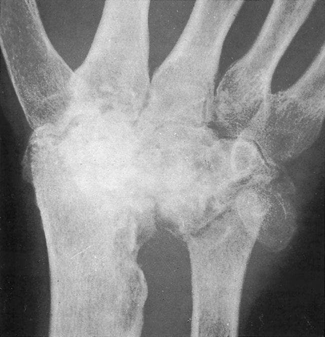
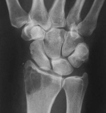
Loss of bone density
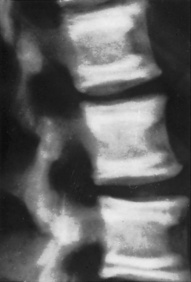
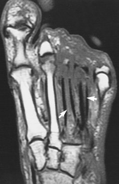
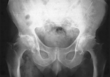
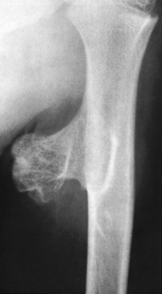
Osteoporosis (Figs 3.1, 3.2)
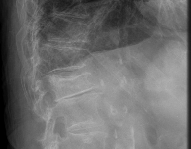
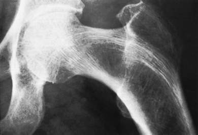
Fig. 3.2 Osteoporosis, femoral head. MR image. Note the joint effusion.
(From Resnick Kransdorf, 2005.)
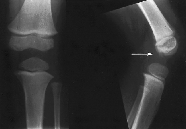
Increase in bone density
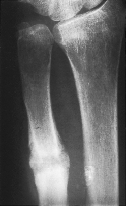
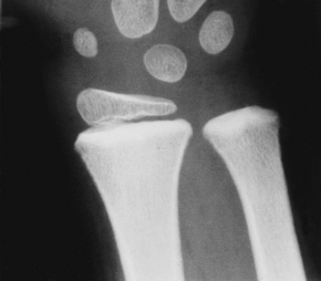
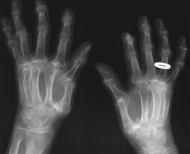
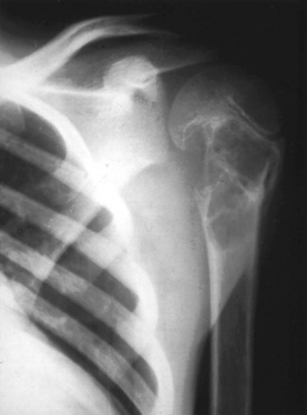
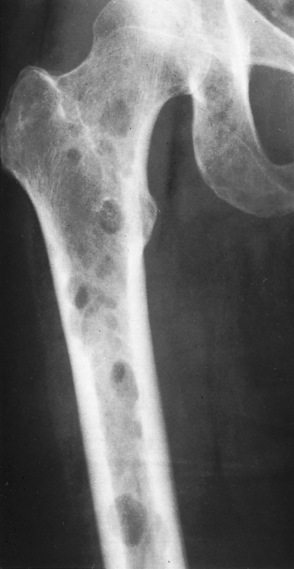
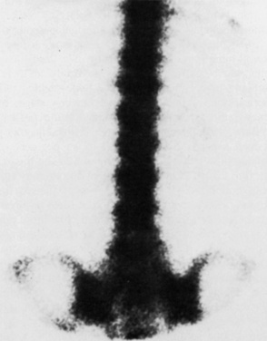
Lead poisoning (Fig. 3.6)
Radiographically dense transverse lines appear at the ends of the shafts of long bones.
Hormone disturbances
Vitamin deficiencies
Vitamins C and D are responsible for the formation of healthy bone tissue.
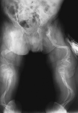
Joint disorders

Bone tumours
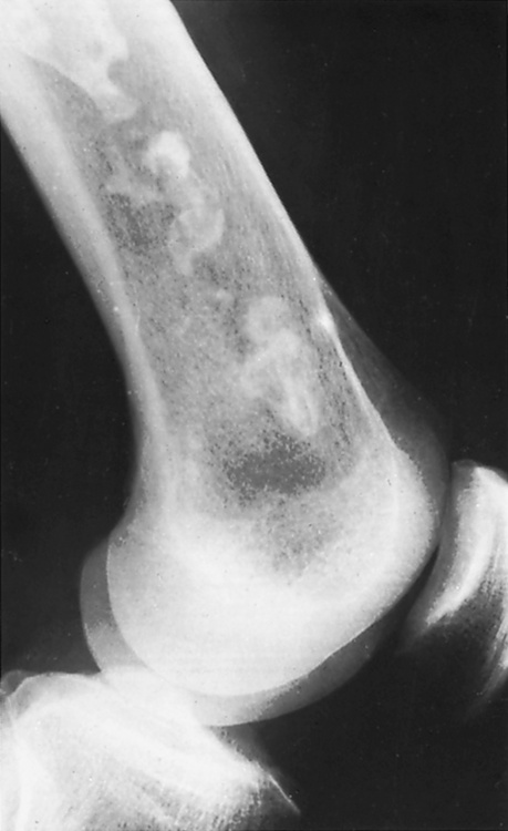
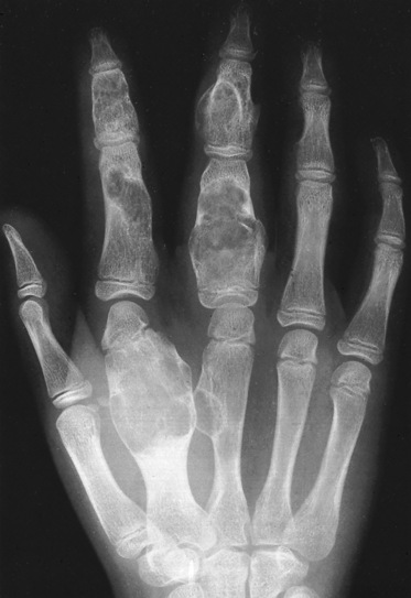
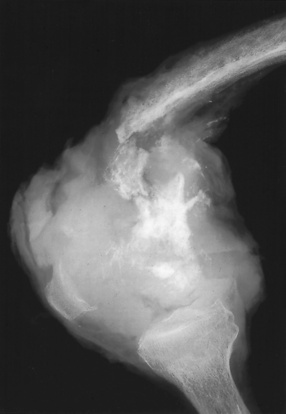
Benign tumours
Radiographically, these have well defined edges and the bone cortex is intact.
Malignant tumours
Fractures
A fracture is an abnormal break in bone continuity and may be complete or partial.
Types of fracture
Simple (closed) fracture
This is where the skin surface remains intact and therefore there is no risk of infection.
Orthopaedic management of fractures


Reduction
Immobilisation
Stages of healing of fractures
1. A blood clot is formed owing to damaged blood vessels in the medulla, cortex and periosteum.
2. Within 24 hours the haematoma is converted into vascular, fibroblastic granulation tissue.
3. After about 7 days cartilage and osteoid tissue are laid down by the osteoblasts, therefore forming irregular, new bone called provisional callus.
4. Provisional callus is converted into ‘normal’ bone containing haversian systems.
5. After a period of time the bone is moulded by the osteoclasts and osteoblasts to regain its original shape.
[/level-membership-for-orthopaedics-category][not-level-membership-for-orthopaedics-category]
3 Pathology
Changes due to pathology
Loss of bone density
Osteoporosis (Figs 3.1, 3.2)
Fig. 3.2 Osteoporosis, femoral head. MR image. Note the joint effusion.
(From Resnick Kransdorf, 2005.)
Increase in bone density
Lead poisoning (Fig. 3.6)
Radiographically dense transverse lines appear at the ends of the shafts of long bones.
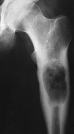
Bone destruction
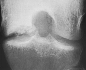
Necrosis of the femoral head following fracture
‘Bone death’ due to a poor blood supply. Radiographically the bone surfaces appear irregular.
