Cardiac cirrhosis: Flattening of Doppler wave form in hepatic veins
“To and fro” motion in hepatic veins and IVC
TOP DIFFERENTIAL DIAGNOSES
• Budd-Chiari syndrome
• Hepatic cirrhosis
• Acute viral hepatitis
CLINICAL ISSUES
• Passive hepatic congestion usually secondary to
Congestive heart failure
Constrictive pericarditis
Tricuspid insufficiency
Right heart failure
• Radiologists may be 1st to recognize cardiac source of liver disease
• Diagnosis is based on clinical and imaging findings
DIAGNOSTIC CHECKLIST
• Differentiate acute passive hepatic congestion from Budd-Chiari and viral hepatitis
• Distinguish chronic, cardiac cirrhosis from other etiologies
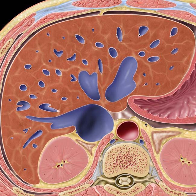
(Left) Graphic shows massive diffuse dilatation of the hepatic veins and mildly heterogeneous liver parenchyma due to passive congestion of the liver.
(Right) Axial CECT in the arterial phase shows early retrograde opacification of dilated hepatic veins and the inferior vena cava (IVC) due to reflux of injected contrast medium through the heart, a sign of impaired antegrade hepatic venous drainage.
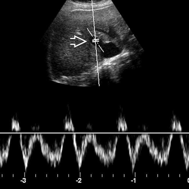
(Left) Increased pulsatility of portal vein Doppler signal is demonstrated in this patient with passive hepatic congestion secondary to tricuspid insufficiency.
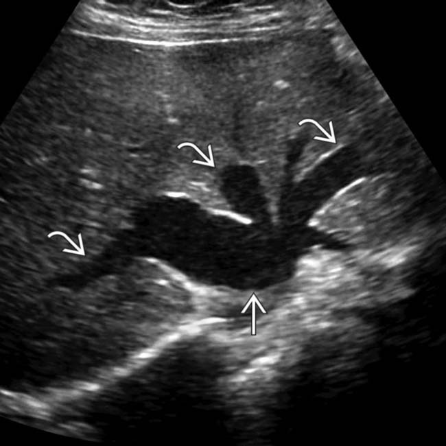
(Right) Transverse ultrasound shows dilated hepatic veins and IVC in a patient with passive hepatic congestion.
TERMINOLOGY
Synonyms
• Congested liver in cardiac disease
Definitions
• Stasis of blood within liver parenchyma as result of impaired hepatic venous drainage
IMAGING
General Features
• Best diagnostic clue
Dilated hepatic veins with “to and fro” blood flow on color Doppler US
Enlarged and tender liver, RUQ pain due to stretched liver capsule
Positive hepatojugular reflux
Pulsatile liver in acute phase
Splenomegaly in late phase
Hepatic failure may be diagnosed before cardiac disease
– e.g., may be misdiagnosed as “cryptogenic cirrhosis” when due to constrictive pericarditis
Lab data
– Acute: Mild abnormal liver function test (LFT)
– Chronic: Grossly abnormal LFT
Diagnosis based on clinical and imaging findings
• Clinical profile
Cardiac patient with hepatomegaly and hepatojugular reflux
Demographics
• Age
Any age group
• Gender
M = F
Natural History & Prognosis
• Complications: hepatic &/or cardiac failure
• Prognosis: good for acute congestion
Chronic phase: Poor
Treatment
• Acute or early phase
Full recovery once patient’s cardiac disease is corrected
• Chronic or late phase
Cardiac cirrhosis may be irreversible, even with correction of cardiac function
DIAGNOSTIC CHECKLIST
Consider
• Differentiate acute passive hepatic congestion from Budd-Chiari and viral hepatitis
• Distinguish chronic, cardiac cirrhosis from other etiologies
Image Interpretation Pearls
• Inferior vena cava and hepatic veins
Dilated and early enhancement (due to reflux)
“To-and-fro” motion on color Doppler
Loss of normal triphasic velocity flow pattern
(Left) Oblique transabdominal ultrasound of a 63-year-old woman with RUQ pain shows circumferential gallbladder wall edema and shadowing posterior to a gallstone .
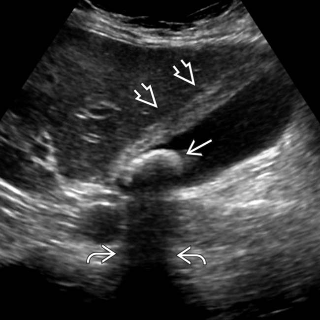
(Right) Axial US image of the hepatic confluence in the same case shows marked dilatation of the retrohepatic IVC and hepatic veins .
(Left) In the same case, Doppler interrogation of the right hepatic vein shows markedly phasic flow.
(Right) Doppler of the main portal vein in the same case shows pulsatile portal venous flow. US findings of dilated IVC and hepatic veins (HVs), gallbladder wall edema, markedly phasic HV flow, and pulsatile portal venous flow are classic findings that indicate passive hepatic congestion and are key in distinguishing this (rather than acute cholecystitis) as the diagnosis.
(Left) Arterial phase CECT in a 57-year-old man with recent myocardial infarction shows marked reflux of contrast material down into the dilated HVs and IVC .
(Right) Portal venous CT in the same case shows HVs lower in the liver are not yet opacified by antegrade flow through the liver. The liver is enlarged and parenchymal enhancement is diminished and very heterogeneous, sometimes described as a “nutmeg liver.”
(Left) Axial arterial phase CECT of a 63-year-old woman initially diagnosed with “cryptogenic cirrhosis” shows reflux of contrast material into dilated HVs and the IVC . A right-sided pleural effusion is also present.
(Right) Arterial phase CECT section in the same case shows heavier, contrast-opacified blood settling within the dependent position of the dilated IVC and even the right renal vein .
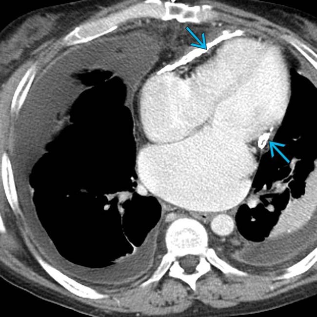
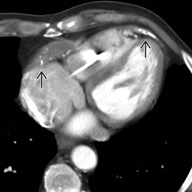
(Left) Axial venous phase CECT in the same case shows bilateral pleural effusions and a thickened, calcified pericardium that compresses and distorts both the right and left ventricles, consistent with constrictive pericarditis.
(Right) Venous phase CECT in the same case shows dilation of the IVC . The liver is diminished in size with a nodular surface.
(Left) Venous phase CECT in the same case shows layering of contrast-opacified blood within the dilated IVC, indicating severe restriction of venous return to the heart.
(Right) Venous phase CECT in the same case shows a nodular hepatic surface and widened fissures. This patient had constrictive pericarditis suggested for the first time based on CT interpretation, which was subsequently confirmed. The chronic passive congestion of the liver had resulted in cardiac cirrhosis.
Axial CECT in a patient with chronic constrictive pericarditis shows soft tissue and calcified thickening of the pericardium as well as deviation of the interventricular septum.
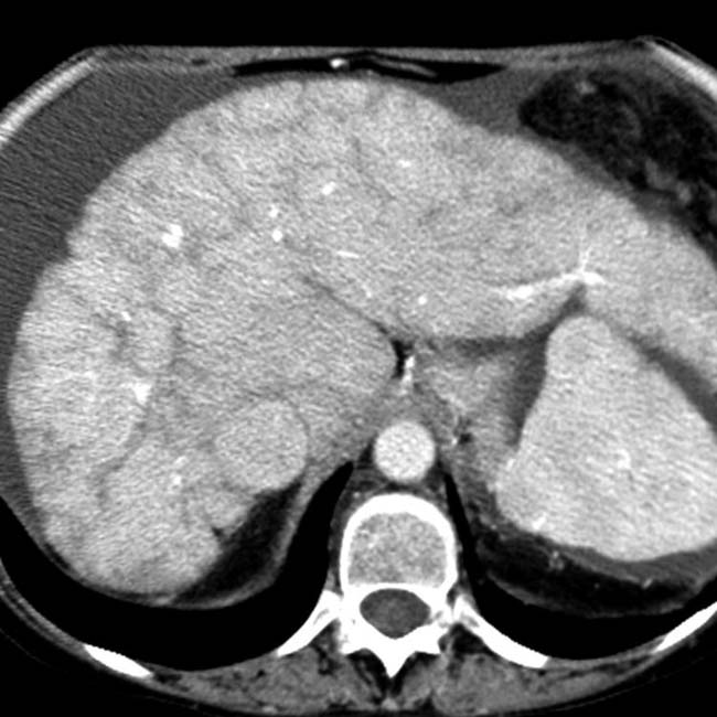
Axial portal venous phase CECT shows mottled enhancement of the liver and a halo of lymphedema around the IVC. This patient presented with passive hepatic congestion secondary to constrictive pericarditis.
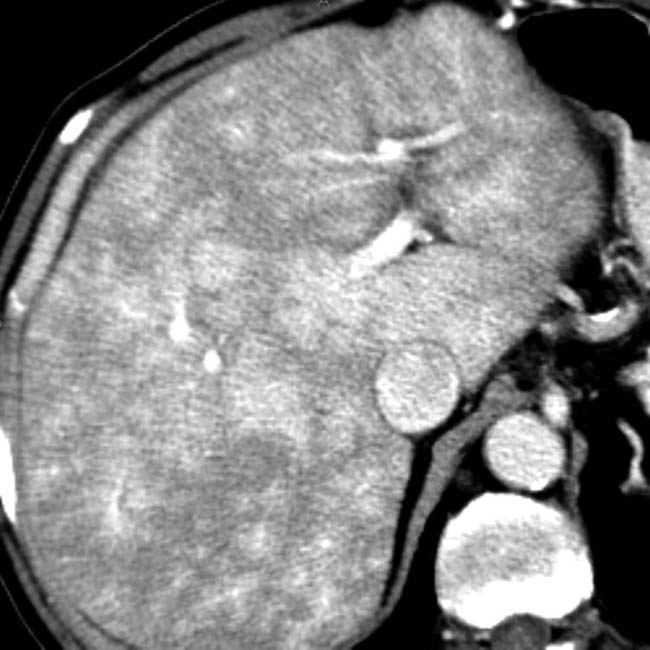
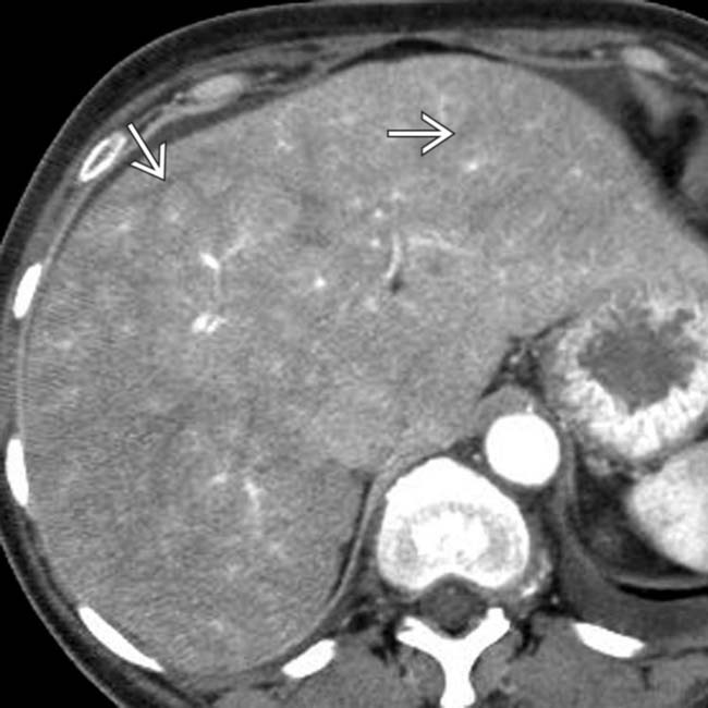
Axial CECT shows typical changes from cardiac cirrhosis. The liver is small and dysmorphic with heterogeneous enhancement.
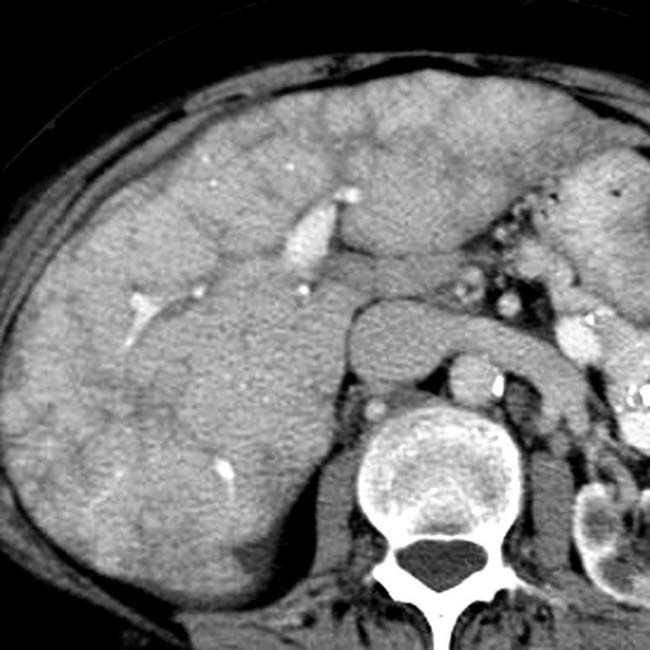
Axial CECT shows a dysmorphic liver with an atrophic right lobe, a hypertrophied lateral segment, and heterogeneous enhancement. Note the ascites.


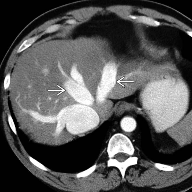
 and the inferior vena cava (IVC) due to reflux of injected contrast medium through the heart, a sign of impaired antegrade hepatic venous drainage.
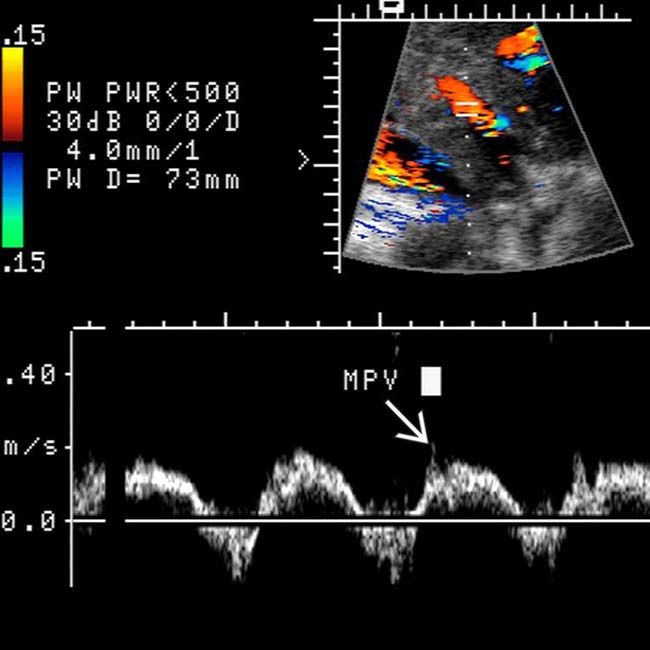
 is demonstrated in this patient with passive hepatic congestion secondary to tricuspid insufficiency.
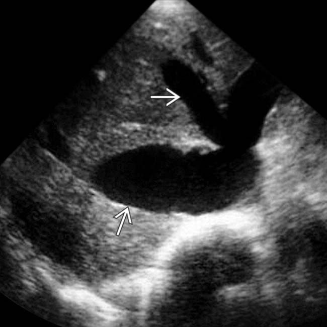
 and IVC in a patient with passive hepatic congestion.


 and shadowing
  posterior to a gallstone
  .
.
 and hepatic veins
  .
.
 shows markedly phasic flow.
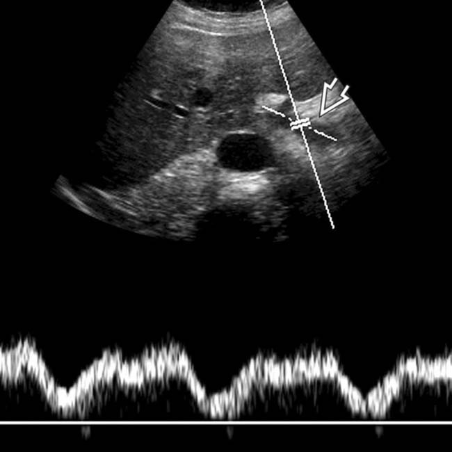
 in the same case shows pulsatile portal venous flow. US findings of dilated IVC and hepatic veins (HVs), gallbladder wall edema, markedly phasic HV flow, and pulsatile portal venous flow are classic findings that indicate passive hepatic congestion and are key in distinguishing this (rather than acute cholecystitis) as the diagnosis.
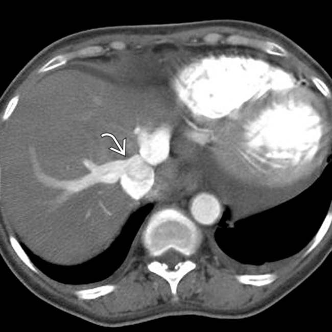
 .
.
 are not yet opacified by antegrade flow through the liver. The liver is enlarged and parenchymal enhancement is diminished and very heterogeneous, sometimes described as a “nutmeg liver.”
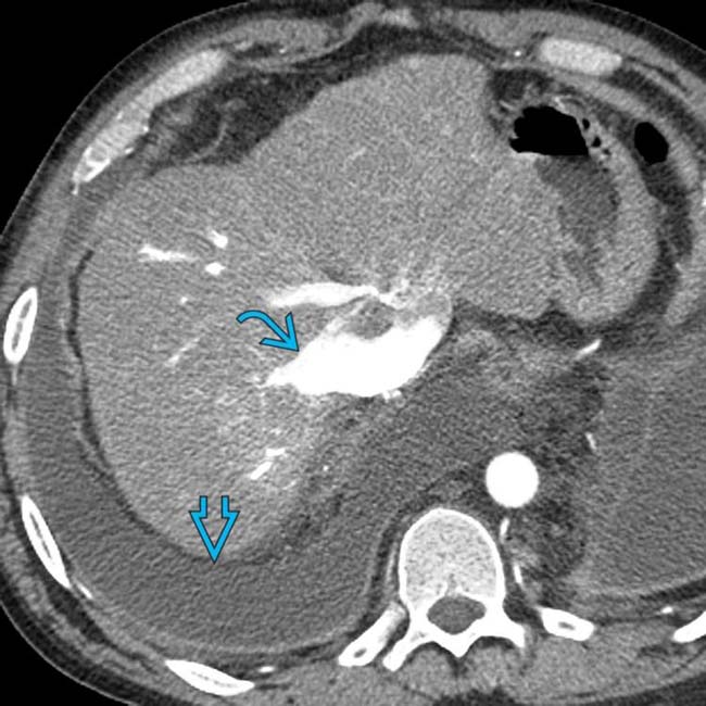
 . A right-sided pleural effusion
  is also present.
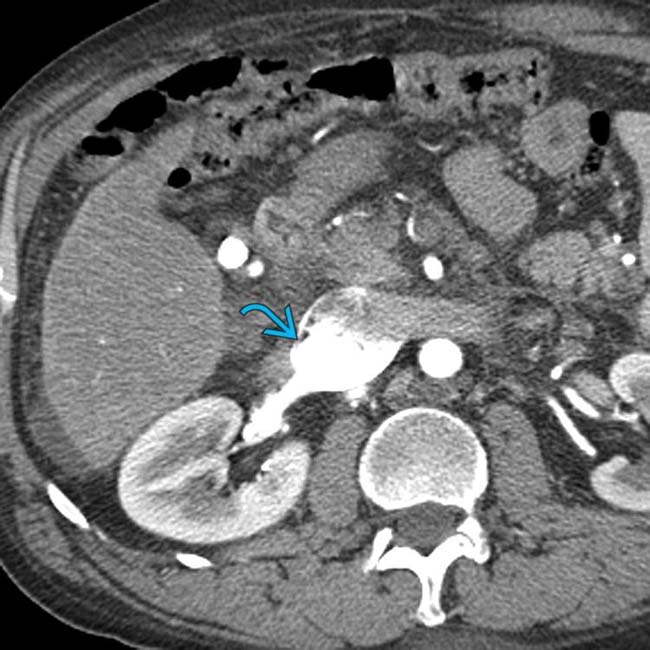
 .
.
 that compresses and distorts both the right and left ventricles, consistent with constrictive pericarditis.
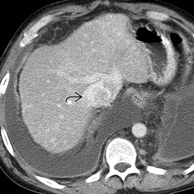
 . The liver is diminished in size with a nodular surface.
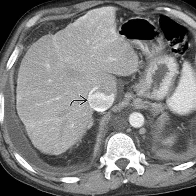
 within the dilated IVC, indicating severe restriction of venous return to the heart.
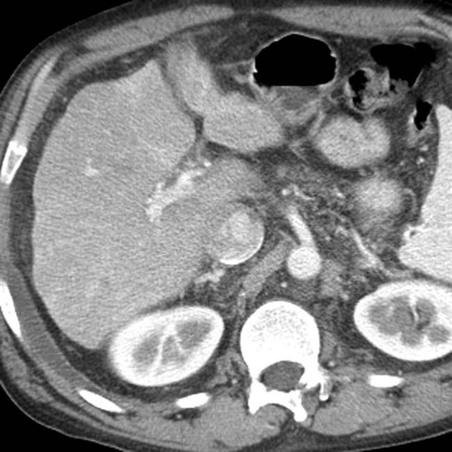
 as well as deviation of the interventricular septum.