Partial Gastrectomy: Bilroth Procedures

Published on 19/07/2015 by admin

Filed under Radiology

Last modified 22/04/2025

Print this page

rate 1 star rate 2 star rate 3 star rate 4 star rate 5 star
Your rating: none, Average: 0 (0 votes)

This article have been viewed 3648 times

 Antrectomy with gastroduodenostomy

• Billroth 2 (B2) procedure

image Distal gastrectomy with gastrojejunostomy
• Surgery for gastric cancer

image May be some variant of B1 or B2, up to total gastrectomy
• Complications include recurrent tumor and acute or chronic sequelae of surgery
• Recurrent or new carcinoma

image Local, lymph node, peritoneal, hematogenous
• Bezoar formation

image Conforms to shape of stomach, traps air within
• Anastomotic leak

image CT may detect indirect signs of leaks missed on upper gastrointestinal (GI) series (up to 50% of cases)
• Duodenal stump leakage

image Loculated collection of fluid in subhepatic space
image Rarely diagnosed on upper GI
• Jejunogastric intussusception

image Rare complication of B2 procedure
• Afferent loop syndrome

image Obstruction of afferent loop at or near anastomosis → dilation of duodenum

DIAGNOSTIC CHECKLIST

• Upper GI series is 1st-line test for detecting mechanical complications of gastric surgery
• CT is optimal test for general surveillance for postoperative complications
• PET/CT is optimal imaging test for surveillance of recurrent gastric carcinoma
• Abscessogram may identify leak as source of infection
image
(Left) Graphic depicts an isoperistaltic Billroth 2 gastrojejunostomy. The afferent limb image
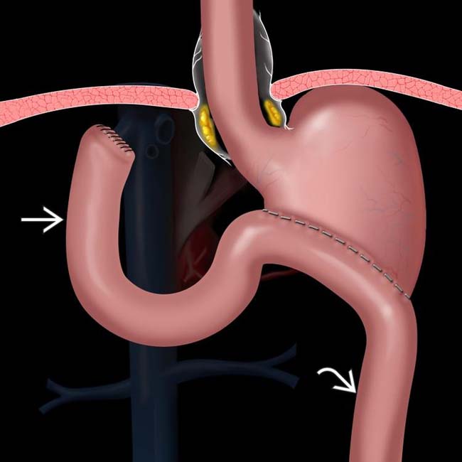
, composed of the duodenum and a variable length of jejunum, carries pancreaticobiliary secretions toward the stomach, while the efferent limb image carries fluid and food downstream.

image
(Right) Graphic depicts an antiperistaltic Billroth 2 procedure, in which the afferent loop image 
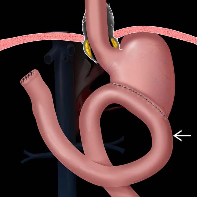
enters the anastomosis from a left-to-right direction. This procedure is intended to reduce the prevalence of bile gastritis.
image
(Left) Film from a small bowel follow-through (SBFT) shows evidence of a prior Billroth 2 procedure and complete obstruction of antegrade flow of barium in the mid jejunum image
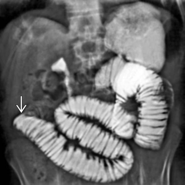
. At surgery, a phytobezoar was removed, which corresponded to the shape and size of the gastric remnant.
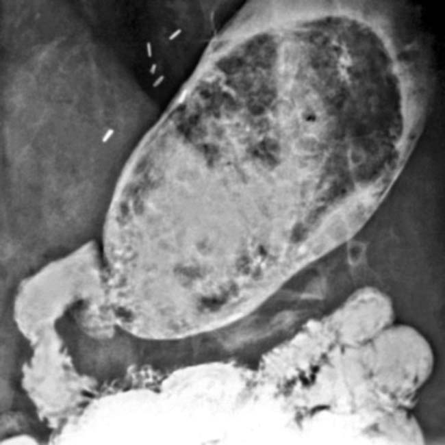
image
(Right) Film from an upper GI series shows evidence of a prior Billroth 1 procedure, along with persistent filling defects within the stomach that conform to the shape of the stomach, a bezoar.

IMAGING

General Features

• Many to most fluoroscopic exams of esophagus, stomach, and duodenum are now performed for patients who have surgically altered anatomy
• Some procedures are so common they are discussed separately

image Postoperative state, esophagus

– Includes esophagectomy with gastric pull-through
image Fundoplication complications
image Bariatric surgery
• Goal for evaluating remaining procedures

image Define expected postoperative anatomy
image Describe imaging approaches to evaluation of postoperative patients
image Describe imaging and clinical findings for various complications

Surgical Procedures

• Billroth 1 (B1)  procedure

image Antrectomy with gastroduodenostomy
image Polya variation: Entire excised end of gastric stump is used for anastomosis
image Hofmeister: Only a portion (usually greater curvature portion) is used
• Billroth 2 (B2)  procedure

image Distal gastrectomy with gastrojejunostomy

– Stomach may be anastomosed to Roux limb or loop of jejunum
– Anastomosis is side to side
– Variable length of duodenum and jejunum forms proximal or afferent loop

image Carries pancreaticobiliary secretions toward stomach
– Distal or efferent loop carries food and fluid downstream
– Isoperistaltic anastomosis (right to left)

image Afferent limb 1st contacts lesser curve side of anastomosis; efferent limb contacts distal side
– Antiperistaltic (left to right)

image Opposite configuration
– Duodenal stump is closed by sutures

image Usually filled with gas bubble on postoperative CT
image Located in subhepatic space
– Anastomotic loop may be brought to gastric remnant in antecolic or retrocolic position

image Antecolic: Results in longer afferent loop
image Retrocolic: Shorter afferent loop; considered more “physiological” and usually preferred
• Surgery for gastric cancer

image Varies according to site and size of tumor
image May be some variant of B1 or B2, up to total gastrectomy

– Gastrectomy usually results in creation of esophagojejunal anastomosis
image Usually involves extensive denervation of stomach

– Alters gastric and intestinal motility and absorption

Complications

• Recurrent or new carcinoma

image Patients who have had partial gastrectomy for gastric cancer have high risk of recurrent tumor

– May occur in gastric remnant or anastomosis (often causes bowel thickening or obstruction)
– Lymph node metastases (celiac, retroperitoneal, mesenteric)
– Peritoneal (nodular thickening of peritoneum or omentum; loculated ascites)
– Hematogenous (liver, lungs, adrenals, ovaries [Krukenberg tumors])
image Patients who have had B2 surgery for benign ulcer disease also are at ↑ risk

– 3-6x ↑ risk of cancer in gastric stump, with 15-20 year latent period
– Related to chronic bile gastritis and achlorhydria
• Gastric stasis

image → postprandial bloating, vomiting, weight loss
image Not always due to mechanical obstruction

– May be due to vagus denervation, gastritis, etc.
• Dumping syndrome

image Symptoms: Weakness, dizziness, sweating, nausea, colic, diarrhea
image Urgent desire to lie down after eating
image Attributed to vasomotor and cardiovascular etiology
image Prevalence of 5-50%, depending on type of procedure
image Usually improves with dietary alterations

– Rarely improved by reoperation
• Bezoar formation

image ↑ prevalence after B1 or B2
image Predisposing factors: Achlorhydria, denervation, edentulous patient, anastomotic stricture
image Distinguish from semisolid food (which will eventually pass through stoma)

– Bezoar conforms to shape of stomach, traps air within, floats on fluid in stomach, does not exit with food
• Anastomotic leak

image Can occur after any procedure

– Highest after surgery for gastric cancer, especially total gastrectomy (with esophagojejunostomy)
– Multifactorial, including debilitated condition
image Early complication

– Days to weeks
image Diagnosed by extraluminal gas, contrast material, fluid

– Upper gastrointestinal (GI) series best for some early leaks
– CT may be necessary for evaluation of complete extent of fluid collection or abscess
– CT may detect indirect signs of leaks missed on upper GI series (up to 50% of cases)
– Loculated collections of fluid and gas, extravasated contrast medium
• Duodenal stump leakage

image Loculated collection of fluid in subhepatic space on CT

– Rarely diagnosed on upper GI
image Bile and pancreatic juice cause peritoneal inflammation (thick and enhancing)
• Stomal ulceration

image May occur after B1 or B2
image Usually on small bowel side of anastomosis
image Difficult to depict ulcer on imaging (due to distorted anatomy, thick folds)

– Endoscopy is preferred technique
• Jejunogastric intussusception

image Rare complication of B2 procedure
image Due to prolapse of jejunum through anastomosis into gastric remnant
image Can cause bowel obstruction ± ischemia

– Can be recognized by upper GI or CT as filling defect within stomach, having jejunal fold pattern, peristalsis
• Afferent loop syndrome

image Obstruction of afferent loop at or near anastomosis → dilation
image Loop distends with pancreatic and bilious secretions
image Symptoms: Pain, nausea, signs of biliary obstruction
image May be missed or indirectly suggested by upper GI

– Afferent loop fails to fill with ingested contrast medium (can be normal)
image CT shows distended afferent limb (2nd and 3rd portions of duodenum)

– May depict tumor in gastric remnant or bowel as obstruction etiology
• Chronic remnant gastritis

image Chronic bathing of gastric mucosa by alkaline bile and pancreatic juice

– Routinely causes gastritis
– May progress to intestinal metaplasia or cancer
– Upper GI and CT show thickened rugal folds ± ulcers or tumor

Imaging Recommendations

• Protocol advice

image Immediate postoperative period

– Water-soluble contrast agents for fluoroscopic upper GI (or CT)

image Use nonionic (low osmolar) agent if aspiration is a concern
– For evaluation of anastomotic leak: Staple line dehiscence, bowel perforation, abscess
– Precede with scout film of abdomen

image Assists in recognizing surgical clips, drains, etc.
image Makes identification of leak more apparent on post-procedure films
image Later evaluations

– Barium is used for upper GI series
– Single contrast study answers most clinically relevant questions
– Double contrast study better shows superficial ulcers but is more difficult to perform and interpret

CLINICAL ISSUES

Presentation

• Other signs/symptoms

image Other complications

– Infection

image Most common source of morbidity and mortality following gastric surgery
image Pulmonary, urinary
image Operative wound; often indicative of anastomotic leak
image Undrained fluid may become infected (CT: Enhancing wall ± gas)
image Pneumoperitoneum may be normal postoperative finding or indicative of leak
image CT may aid in image-guided drainage procedures
– Esophageal dysmotility

image Effects of vagotomy or surgical injury
– Esophagitis

image From acid or alkaline reflux
– Gastric-emptying problems

image Gastroparesis or outlet obstruction
image Bezoar
image Generalized ileus
image Fistulas (gastrocolic, gastrojejunal)
– Malabsorption

image Inadequate mixing of pancreatic juice, bile, enteric contents
image Inadvertent gastroileostomy
– Hemorrhage

image Sentinel clot (heterogeneous high attenuation blood [45-65 HU] near source of bleeding)
– Pancreatitis

image Due to injury of pancreas at surgery or invasion of pancreas by gastric tumor requiring partial pancreatectomy

DIAGNOSTIC CHECKLIST

Consider

• Upper GI series is 1st-line test for detecting mechanical complications of gastric surgery

image Leak, obstruction, etc.
image Misses leaks in up to 50% of cases (in some series)
• CT is optimal test for general surveillance of postoperative complications

image Infection, mechanical problems, tumor, etc.
• PET/CT is optimal imaging test for surveillance of recurrent gastric carcinoma
• Abscessogram

image Injection of contrast medium through surgically or radiologically placed drains
image May identify opacification of bowel or stomach

– Indicates leak as source of infected fluid collection

Image Interpretation Pearls

• Try to identify anatomy on CT and use medical records (op-reports, etc.)

image Important to identify, for instance, what constitutes the afferent limb

– Always in continuity with 3rd portion of duodenum
– Crosses between aorta and superior mesenteric vessels

image
(Left) 30-minute delayed film from a SBFT in an elderly man, who had a Billroth 2 procedure for benign ulcer disease 15 years prior, shows preferential filling of the afferent limb image 
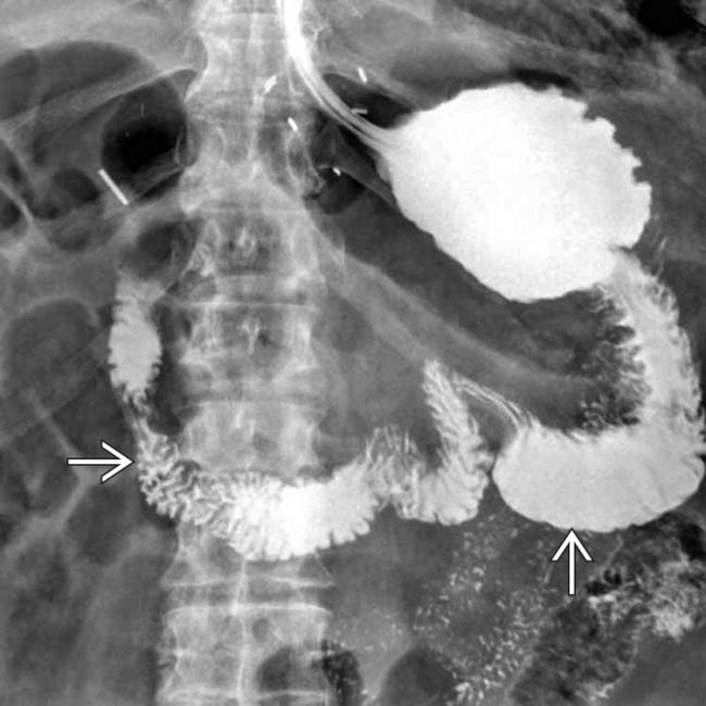
and delayed and decreased filling of the efferent limb.
image
(Right) Axial CECT in the same patient shows luminal distention and wall thickening of the gastric remnant image 
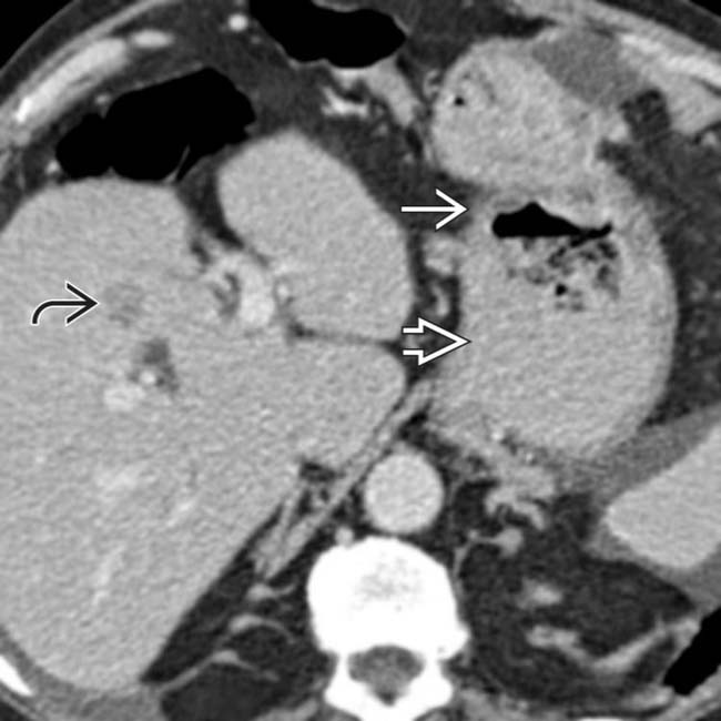
near the gastroenteric anastomosis image, due to gastric carcinoma. Liver metastases are evident image.
image
(Left) Axial CECT in the same patient shows extensive mesenteric lymphadenopathy image 
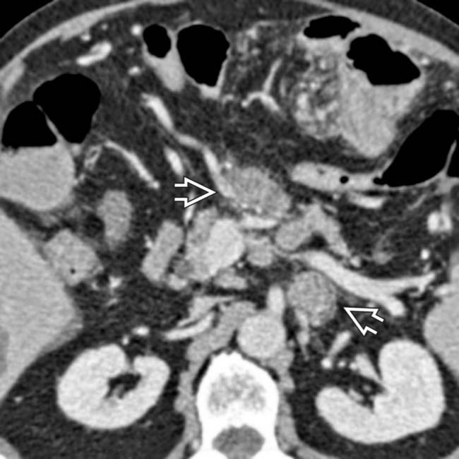
from lymphatic metastases.

image
(Right) Axial CECT in the same patient shows a large mesenteric tumor encasing the efferent limb image
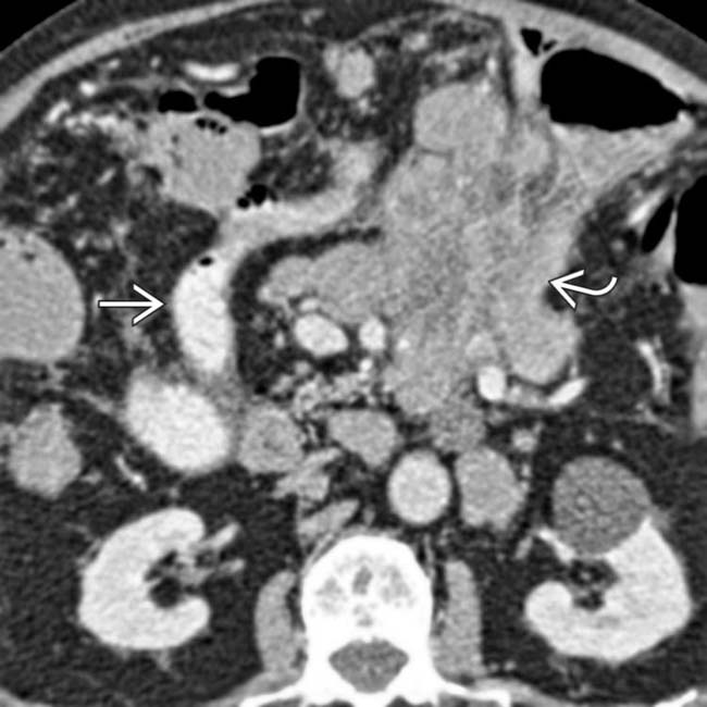
. The contrast-filled lumen of the afferent limb is seen image. Gastric cancer occurs with increased frequency following this type of ulcer surgery, usually 15 or more years later.
image
(Left) Jejunogastric intussusception as a complication of partial gastrectomy with Billroth 2 anastomosis. CT demonstrates a loop of thick-walled and ischemic jejunum image 
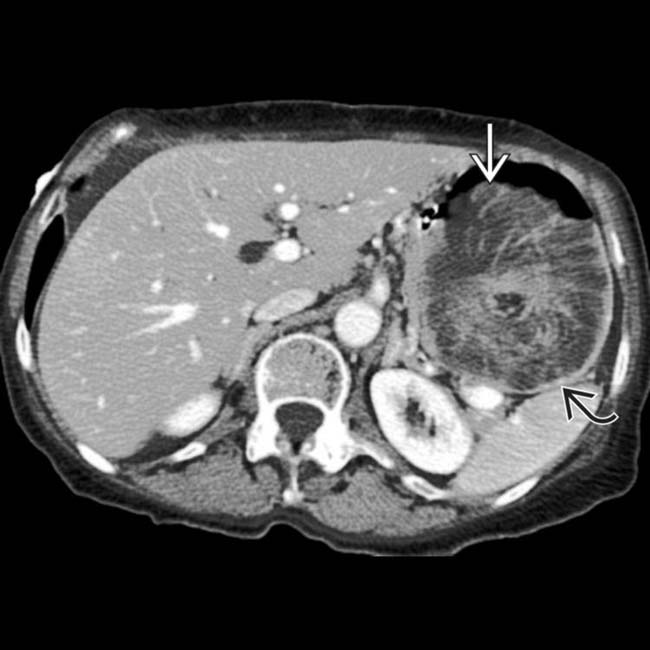
within the distended gastric remnant image.

image
(Right) Coronal reformation CECT in the same patient shows the retrograde intussusception image 
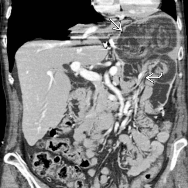
with invagination of bowel, mesenteric fat, and vessels image into the gastric remnant (intussuscipiens). This is a rare complication but may result in bowel obstruction and ischemia.

Share this: