Papulosquamous eruptions
Papulosquamous eruptions are raised, scaly and marginated, and include psoriasis, lichen planus and other conditions listed in Table 1. Eczema is not included as it does not usually have a sharp edge. These eruptions are not related aetiologically. Several are characterized by fine scaling and have the prefix ‘pityriasis’, which means ‘bran-like scale’.
Table 1 Papulosquamous eruptions
Pityriasis rosea
Clinical presentation
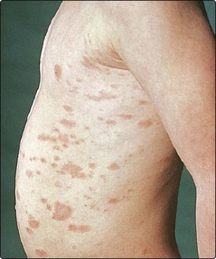
The generalized eruption is preceded in most patients by the appearance of a single lesion, 2–5 cm in diameter, known as a ‘herald patch’ (Fig. 1). Some days later, many smaller plaques appear, mainly on the trunk but also on the upper arms and thighs. Individual plaques are oval, pink and have a delicate peripheral ‘collarette’ of scale. They are distributed parallel to the lines of the ribs, radiating away from the spine. Itching is mild or moderate. The eruption fades spontaneously in 4–8 weeks. It tends to affect teenagers and young adults. The cause is unknown, but epidemiological evidence of ‘clustering’ suggests an infective aetiology.
Pityriasis (tinea) versicolor
Clinical presentation
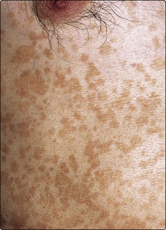
The condition is caused by overgrowth of the mycelial form of the commensal yeast Malassezia (previously Pityrosporum ovale) and is particularly common in humid or tropical conditions. In Europe, it mainly affects young adults, appearing on the trunk and proximal parts of the limbs (Fig. 2). In untanned, white caucasians, brown or pinkish oval or round superficially scaly patches are seen, but, in tanned or racially pigmented skin, hypopigmentation is found as a result of the release by the organism of dicarboxylic acids that inhibit melanogenesis.
Reiter’s disease
Reiter’s disease is a syndrome of polyarthropathy, urethritis, iritis and a psoriasiform eruption.
Clinical presentation and management
Reiter’s disease almost invariably affects males who have the HLA-B27 genotype and commonly follows a genitourinary or bowel infection. The joint and eye changes are often severe. Skin involvement includes a balanitis (p. 121) and red, scaly, pustular, psoriasiform plaques on the feet (keratoderma blenorrhagicum).
Chronic superficial dermatitis
Previously known as parapsoriasis, a term best avoided, this is an uncommon chronic dermatitis of small scaly pink–brown oval or round-shaped plaques, mainly on the trunk. The variant with larger plaques may proceed to mycosis fungoides (cutaneous T cell lymphoma) or be this from the onset.
Clinical presentation
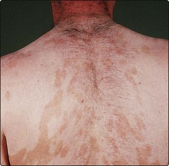
In chronic superficial dermatitis, scaly patches develop, usually on the abdomen, buttocks or thighs (Fig. 3). The onset is in young to mid-adult life, and the plaques are indolent. It may be difficult to predict which cases will progress to mycosis fungoides (p. 104), especially as the evolution may take place over many years, but the ‘benign’ lesions tend to be small and finger-like in shape, whereas the ‘premalignant’ plaques are larger, asymmetrical, atrophic and can show associated poikiloderma (reticulate pigmentation, telangiectasia and atrophy). Biopsy is necessary to look for the changes of mycosis fungoides, and further biopsy of any changed area is required. The disease is often indolent and may persist over a period of several years.
Other pityriases
Other varieties of pityriasis include the following:
 Pityriasis lichenoides. A rare chronic eruption in which small papules topped by a fine single scale appear on the limbs and trunk. It is seen in adolescents and young adults and may occur in an acute form (Fig. 4), which heals with scarring.
 Pityriasis rubra pilaris. A rare, scaly follicular eruption, which may progress to erythroderma (see p. 44).

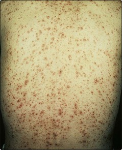
Secondary syphilis
Clinical presentation
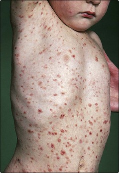
The secondary phase of syphilis (p. 120) starts 4–12 weeks after the appearance of the primary chancre and consists of an eruption, lymphadenopathy and variable malaise. Pink or copper-coloured macules, which later develop into papules, appear in a symmetrical distribution on the trunk and limbs and are non-itchy (Fig. 5). Annular patterns are not uncommon, and involvement of the palms and soles is distinctive. Other signs are moist warty lesions (condyloma lata) in the anogenital area, buccal erosions that may be arcuate (snail-track ulcers) and a diffuse patchy alopecia. Mucosal lesions are infectious. Without treatment, the lesions of secondary syphilis resolve spontaneously in 1–3 months.
Differential diagnosis and management
Pityriasis rosea, psoriasis, drug eruption, infectious mononucleosis, rubella and measles may need to be considered. Treponemal serology is positive in all patients with secondary syphilis. Treatment is with intramuscular benzathine benzylpenicillin (p. 120). Patients with syphilis are best managed by physicians familiar with the treatment of genitourinary infections.


