203 Papilloedema
Questions
Advanced-level questions
Mention a few causes of papilloedema
Mention a few conditions that simulate papilloedema
• Medullated nerve fibres: seen on the disc or even on the retina. The appearance is typically flared and on focusing will reveal fibres traversing the area. Field defects are the result of the retinal vessels being obscured. Since these are present from birth, the patient is unaware of the defect.
• Bergmeister’s papilla: whitish elevation of the centre of the disc with venous and arterial sheathing. It is common and seen at all ages. There is an equal sex and racial incidence.
• Pseudopapilloedema: congenitally elevated discs secondary to hyaloid tissue (drüsen) or hyperopia.
Note: Elevation or swelling on the optic disc occurs in the following conditions:
How do you differentiate papillitis from papilloedema?
| Papillitis | Papilloedema |
|---|---|
| Usually unilateral | Usually bilateral |
| Visual acuity is considerably reduced in relation to the degree of swelling of the disc | Visual acuity only slightly reduced until late stages |
| Visual field defect is usually central, particularly for red and green | Peripheral constriction or enlargement of the blind spot |
| Marcus Gunn pupil may be present | Marcus Gunn pupil is absent |
| Eye movements may be painful | Eye movements are never painful |
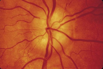
What are the stages of papilloedema?
• Stage I: increase in venous calibre and tortuosity.
• Stage II: optic cup becomes pinker and less distinct, the vessels seeming to disappear suddenly on the surface of the disc.
• Stage III: blurring of the discs on the nasal side. (In many normal discs, the nasal edge is less distinct and one of the most frequent false-positive signs is questionable blurring of the nasal disc margins.)
• Stage IV: the whole disc becomes suffused and slightly elevated. The margins may disappear and the vessels seem to emerge from a mushy swelling. The optic cup is filled and there are haemorrhages around the disc.


