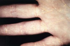
175 Palmar xanthomata
Salient features
Examination
• Yellowish-orange discolorations over the palmar and digital creases (Fig. 175.1 and see Fig. 173.1E)
• Look for the following signs:
• Tell the examiner that this patient probably has a type III hyperlipidaemia.
Note: A more generalized form may be associated with monoclonal gammopathy of myeloma or lymphoma.
Advanced-level questions
How would you classify hyperlipidaemia?
Fredrickson classification, depending on laboratory findings (see Fig. 173.1 for clinical correlates):
• Type I: raised levels of chylomicrons and triglycerides, normal cholesterol concentration (pancreatitis, eruptive xanthomata and lipaemia retinalis)
• Type IIa: raised LDL and cholesterol levels, normal concentration of triglycerides (premature coronary artery disease, tendon xanthomata and arcus corneae)
• Type IIb: raised levels of LDL, VLDL, cholesterol and triglycerides (premature coronary artery disease)
• Type III: raised β-VLDL (cholesterol-rich) remnants, cholesterol and triglycerides (premature coronary artery disease, peripheral vascular disease, palmar and tuberous xanthomata)
• Type IV: raised VLDL and triglycerides, normal cholesterol (premature coronary artery disease: in some forms, risk of developing chylomicronaemia syndrome)
• Type V: raised chylomicrons, VLDL, cholesterol and triglycerides (pancreatitis, eruptive xanthoma, lipaemia retinalis).


