Finger Pain
A painful finger is a common presenting symptom. This may vary from an obvious local lesion to part of a generalised disease, e.g. rheumatoid arthritis. Impairment of the function of even a single finger may grossly impair the overall function of the hand.
Causes
History
Traumatic
There will be a history of trauma, often a crushing injury. Subungual haematoma results from ‘trapping’ of the nail. It is extremely painful, as a bruise comes up between the nail and the nailbed.
Inflammatory/infective
Paronychia
This presents as a painful, tender spot close to the nail. It throbs and may keep the patient awake. There may be a history of picking the skin around the nail. Pus may exude from the side of the nail.
Pulp space infection
This occurs in the pulp space of the fingertip. There may be a history of penetrating injury, e.g. a prick with a sharp object. There is pain, redness and swelling, and the finger throbs.
Neoplastic
Glomus tumour
This is a rare lesion but it is very painful. The patient complains of severe pain every time the nail is touched, the most common site being below the nail.
Primary and secondary bone tumours
These are rare in the phalanges. Pain and swelling occurs and there may be a history of a primary tumour, e.g. breast, bronchus, thyroid, kidney or prostate.
Degenerative
Rheumatoid arthritis
Women are more commonly affected than men. The usual symptoms are pain, swelling and stiffness of the fingers. General malaise may occur. The patient may complain of deformities of the fingers.
Vascular
Chilblains
The simplest vascular problem that affects the fingers is chilblains. Women are more affected than men. The patient complains of a swelling on the side or backs of the fingers, which develops rapidly after exposure to cold. The lesion is painful and itches. The lesions are usually multiple and occur most commonly in winter.
Small vessel disease
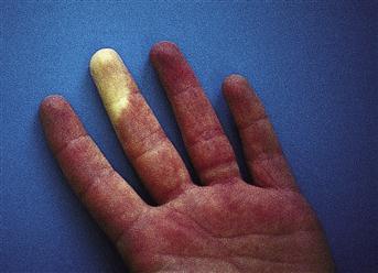
Ischaemia results in pain, discoloration, ulceration or frank gangrene. The patient may complain of sudden onset of a cold, painful finger or frank gangrene may be apparent. There may be a history of Raynaud’s phenomenon, Buerger’s disease, diabetes or scleroderma. There may be cardiac disease, peripheral vascular disease or a cervical rib to suggest embolism. With Raynaud’s phenomenon, there are often the characteristic changes of sudden onset of pallor induced by exposure to the cold, followed by cyanosis, followed by sudden vasodilatation with a painful, tingling, red digit.
Metabolic
Gout
Gout usually affects the first MTP joint but may affect any joint. The patient will complain of sudden onset of pain, swelling and redness in relation to a joint. There may be a previous history of gout. There may be a family history. An attack may be precipitated by trauma, starvation, infection, diuretics, cytotoxic or immunosuppressive drugs. Gouty tophi may be present.
Autoimmune
Scleroderma
Females are affected more than males. There may be thickening of the fingers. The patient may also complain of Raynaud’s phenomenon and splits and ulcers of the fingertips. There may be a change in facial appearance and the patient may complain of dysphagia.
Neurological
Carpal tunnel syndrome
The patient will complain of pain, paraesthesia in the thumb, index and middle fingers. Worse in bed at night. Relieved by hanging the arm out of bed.
Cervical lesions
The patient may complain of pain or tingling in the fingers. There may be a history of cervical spondylosis or other previous problems with the cervical spine.
Examination
Traumatic
Deformity, redness and swelling to suggest a fracture will be obvious. ‘Trapping’ of the nail or a blow to it will cause a collection of blood under the nail. The diagnosis is usually obvious from the history and the patient will be in considerable pain.
Inflammatory/infective
Paronychia
The skin at the base and side of the nail is red, shiny and bulging. A bead of pus may be seen discharging from under the nail.
Pulp space infection
There is swelling and redness over the pulp of the finger. A pus-filled blister may be seen. If the tension is not rapidly relieved, it may either discharge into the tendon sheath or may cause pressure necrosis and osteomyelitis of the distal part of the terminal phalanx.
Neoplastic
Glomus tumours
These are rare. They are angioneuromyomas. If the tumour occurs under the nail (most common site), there is a small purple-red spot below the nail.
Primary and secondary tumours
These are rare. There is palpable bony swelling, which may or may not be tender.
Degenerative
Rheumatoid arthritis
The finger joints enlarge and become fusiform. Joint deformities occur, with ulnar deviation at the wrist and hyperextension of the proximal interphalangeal joints. There is wasting of muscles of the hand.
Vascular
Chilblains
These usually occur on the dorsum and side of the fingers. The skin over the swelling is reddish-blue. The swellings are oedematous and may burst and ulcerate. There are usually chilblains on the ankles and feet as well as on the fingers.
Small vessel disease
In ischaemic patients, there may be pallor, cyanosis or even frank gangrene of a finger. Ischaemic ulcers may be apparent on the fingertips. The pulp of the fingers may be wasted. Palpate for a cervical rib. Check the pulse for atrial fibrillation (emboli).
Metabolic
Gout
Acute inflammation of a joint occurs, the skin being tense, shiny, hot and red over the joint. The presence of gouty tophi locally or elsewhere, e.g. on the ears, suggests the diagnosis.
Autoimmune
Scleroderma
The skin of the hands has a thickened, white, waxy appearance. The fingers appear swollen with thickened skin, while the pulps of the fingers may be wasted. Small subcutaneous nodules, which are hard, may be palpable. These are due to calcinosis. Look for other signs of scleroderma, e.g. the skin of the face looks tight and shiny and is puckered around the mouth. There may be multiple telangiectasias over the face. Evidence of weight loss due to dysphagia may be present.
Neurological
Carpal tunnel syndrome
Wasting of the thenar muscles. The reduction of sensation in distribution of the median nerve in the hand occurring in advanced cases. Pressure over the carpal tunnel may reproduce symptoms.
Cervical spondylosis
Check sensation in the fingers. Check also the range of neck movements and the reflexes in the upper limbs.
General Investigations
■ FBC, ESR
Hb ↓ anaemia of chronic disease, e.g. rheumatoid arthritis. WCC ↑, e.g. tendon sheath infection. ESR ↑, e.g. rheumatoid arthritis.
■ U&Es
Urea and creatinine raised in chronic renal failure, which may be associated with gout or scleroderma.
■ Rheumatoid factor
Rheumatoid arthritis.
■ ECG
Atrial fibrillation – emboli.
■ Hand X-ray
Rheumatoid arthritis. Bone lesions. Pulp space infection – pressure necrosis with osteomyelitis of the distal part of the terminal phalanx. Calcium deposits in connective tissue in CREST syndrome.
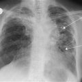
■ CXR with thoracic inlet
Cervical rib – Raynaud’s phenomenon, emboli.
■ Cervical spine X-ray
Cervical spondylosis with referred pain.