Facial Pain
Most patients are able to distinguish between pain arising from the cranium, which is termed headache, and that of the face, which is covered here under the heading of facial pain.
History
In the majority, facial pain originates from local structures. Therefore when formulating a differential diagnosis, consider the conditions affecting the anatomical structures of the face.
Site
The site of pain is perhaps the most useful feature when discriminating between the causes. Although local tenderness may direct the clinician to the affected site, do not forget to consider referred pain from adjacent structures. Pain in the region of the ear may be referred from the skin, teeth, tonsils, pharynx, larynx or neck. Tenderness over the maxilla may be due to sinusitis, dental abscess or carcinoma.
Character
Patients with trigeminal neuralgia often describe paroxysms of sharp, severe pains in the distribution of the trigeminal nerve or major divisions. Pain associated with infections of structures such as the teeth, mastoid and ear often has a dull, aching quality.
Precipitating factors
Pain precipitated by food or chewing action may be due to a dental abscess, salivary gland disorder, salivary duct disorder, temporomandibular joint disorder or jaw claudication due to temporal arteritis. Pain arising from most structures is aggravated by touch; however, with trigeminal neuralgia, even the slightest stroking of the skin is sufficient to produce intense pain (allodynia).
Associated symptoms
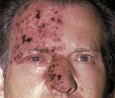
Tears may result when the lacrimal duct is obstructed by nasopharyngeal carcinoma. Complaints of otorrhoea and hearing loss should direct the clinician to infections affecting the ear or mastoid. Nasal obstruction and rhinorrhoea occur with maxillary sinusitis and carcinoma of the maxillary antrum. These may be accompanied by unilateral epistaxis. Swelling of the cheek may be experienced with dental abscesses and maxillary carcinoma. Proximal muscle pain, stiffness and subjective weakness may accompany temporal arteritis due to associated polymyalgia rheumatica. Paraesthesia in the distribution of the trigeminal nerve often precedes the rash of herpes zoster.
Examination
Inspection
A thorough inspection of the face, salivary glands, ear, nose and throat may produce a wealth of information. This involves the use of specific equipment, such as the otoscope and nasal speculum. Unilateral erythema and vesicles in the distribution of the trigeminal nerve may be striking with herpes zoster infection but may not be present in the early stages of the disease. Localised areas of erythema or swelling may also correspond to the site of infection, such as the sinus, ear, mastoid and parotid gland. Post-auricular swelling and downward displacement of the pinna may be observed with mastoiditis. Maxillary swelling and erythema can be caused by dental abscess or maxillary carcinoma. With otitis media, injection or perforation of the tympanic membrane may be visualised with an otoscope. Nasopharyngeal carcinoma may be seen on direct inspection of the nose and throat. Facial paralysis may be seen with infiltration of the facial nerve by tumours of the parotid gland.
Palpation
Gentle stroking of the skin of the face will precipitate severe pain with trigeminal neuralgia. Tenderness may be elicited over the frontal bone with frontal sinusitis, and over the maxilla with maxillary sinusitis. With dental abscesses, pain is elicited over the maxilla or mandible. Mastoid tenderness occurs with mastoiditis or otitis media, and parotid tenderness can result from either mumps or parotitis. Tenderness along the course of the superficial temporal artery is suggestive of temporal arteritis. Movement of the pinna produces pain with otitis externa.
Palpation of the superficial lymph nodes of the neck may reveal lymphadenopathy in the distribution of the lymph drainage of structures affected by infection or carcinoma.
General Investigations
■ FBC
Hb ↓ malignancy. WCC ↑ infection.
■ ESR and CRP
Malignancy and temporal arteritis.
■ X-rays
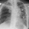
X-rays of the sinuses show mucosal thickening with air-fluid levels in sinusitis. Occasionally total opacification is seen with sinusitis and mastoiditis on mastoid films. In carcinoma of the sinuses, there may be complete opacification of the sinus and destruction of the adjacent bone. CT will reveal the extent of invasion.


