Chest Pain
Chest pain is a common presenting symptom of disorders that can range from trivial to life-threatening.
History
Character
The character of angina is tight and crushing, while the pain from aortic dissection has a tearing quality. Oesophageal reflux may be described as a burning pain, and peptic acid-related pain tends to be deep and gnawing.
Location
The pain from angina and oesophageal reflux may be located retrosternally, and they both can radiate to the jaw or down into the left arm. The pain from pericarditis may be centrally located and radiate to the shoulders (trapezius ridge pain). Pain from aortic dissection often radiates into the back and occasionally into the abdomen (depending on the extent of the dissection). Pulmonary pain can be located anywhere in the thorax.
Precipitating factors
Angina may be precipitated by effort, a defining characteristic. Other known precipitants of angina are emotion, food and cold weather. If angina occurs at rest for more than 20 min, it should be treated as a myocardial infarction until proven otherwise. Oesophageal reflux is often related to meals and precipitated by changes in posture, such as bending or lying. Pain originating from pericarditis and pulmonary origin is often pleuritic, i.e. worse on inspiration; however, musculoskeletal pain can also be worse on breathing due to movement of the thorax.
Relieving factors
Both oesophageal spasm and angina may be relieved by GTN, which relaxes smooth muscle. Antacids will relieve the pain of oesophageal reflux but not angina. The pain associated with pericarditis may be relieved by sitting forwards.
Trauma
A history of a blunt or stretching injury immediately suggests the underlying aetiology of chest wall tenderness and it is important to diagnose rib fractures that can result from more severe trauma. Pathological rib fractures that result from minor injuries may be due to bony metastasis or osteoporosis.
Examination
Temperature
Pyrexia can occur with pneumonia, myocardial infarction, pericarditis and herpes zoster infection.
Pulse
Heart rate on its own is not discriminating, as pain invariably leads to tachycardia. However, palpating both upper and lower limb pulses may be useful. Occasionally peripheral pulses are absent in patients with aortic dissection.
JVP
The JVP is elevated with congestive cardiac failure and acute right ventricular failure, an occasional complication of inferior myocardial infarction and pulmonary embolism (when more than 60% of the pulmonary vascular supply is occluded).
Palpation of the chest
Chest wall tenderness would imply a musculoskeletal cause. The presence of unilateral tenderness confined to a single or adjacent group of dermatomes would suggest either central (vertebral or spinal origin) or peripheral nerve pathology (herpes zoster infection). The trachea deviates away from the side of a tension pneumothorax, and chest expansion is decreased on the same side for pneumonia and pneumothorax. Dullness to percussion will be noted in an area of consolidation with pneumonia and hyperresonance with pneumothorax (a difficult sign to interpret).
Auscultation of the chest
The unilateral absence of breath sounds is consistent with a pneumothorax; more localised loss occurs over an effusion. Bronchial breath sounds are audible over a segment of consolidation and sometimes above the level of an effusion. Localised areas of crepitations are audible with lobar pneumonia, whereas widespread crepitations suggest multilobar involvement or pulmonary oedema secondary to left ventricular failure (from myocardial infarction).
A friction rub may be auscultated with both pericardial and pleuritic disease, but only a pericardial rub will be present when the patient holds the breath.
General Investigations
■ FBC
An elevated white cell count will be expected with pneumonia and, to a lesser extent, in a myocardial infarction.
■ Serum cardiac markers
Following a myocardial infarction, cardiac troponin rises within six hours and remains elevated for up to two weeks.
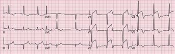
■ ECG (Fig. 9)
Angina or a myocardial infarct will result in ECG changes following the anatomical pattern of coronary vascular insufficiency. Involvement of the left main coronary artery would result in ECG changes of ischaemia or infarction in the anterior (V3–4) and left lateral (V4–6) leads. Involvement of the left anterior descending artery will be reflected in the anterior leads, and insufficiency of the posterior descending artery will produce changes in the inferior leads (II, III, aVF). Changes suggestive of ischaemia or NSTEMI are ST segment depression and T wave inversion. Elevated serum cardiac markers distinguish NSTEMI from angina. ST segment elevation infarcts (STEMI), by definition, produce ST segment elevation and Q wave formation in the corresponding leads. Pulmonary emboli usually result in non-specific ECG changes, such as tachycardia, right axis deviation, right ventricular strain and atrial fibrillation. Occasionally one may find the S1Q3T3 pattern (S wave in lead I, Q wave and an inverted T wave in lead III), indicative of right heart strain.
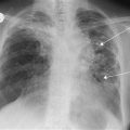
■ CXR
The line of the pleura with absence of lung markings distal to it is the usual finding for a pneumothorax. Areas of consolidation on a chest film may be confined to a lobe or widespread in bronchopneumonia. The classic wedge-shaped shadow (base is distal) of a pulmonary embolus is rare, and occurs only if pulmonary infarction resulted. Dissection of the aorta may widen its width, causing a bulge to appear at the right mediastinal border. Rib fractures or secondary deposits in ribs may be visible.
Specific Investigations
■ V/Q scan
Will show a mismatch in the majority of pulmonary emboli.
■ Pulmonary angiography
Pulmonary emboli will be delineated. It is possible to visualise the site and extent of the embolism and it may also be possible to extract the emboli using a catheter.
■ CT aortography
Confirm and assess the extent and site of the dissection of the aorta.