132
Paget disease of the breast
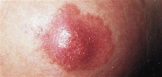
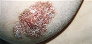
Early Paget disease. There is erythema and scale in the center of the nipple. The areola is not involved.
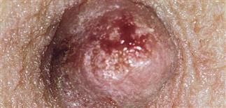
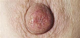
The disease begins insidiously in one breast with a small area of erythema on the nipple that drains serous fluid and may form a crust.
Malignant cells migrate through the epidermis, and the disease becomes initially apparent on the areola and much later on the surrounding skin.
The process appears eczematous, but the plaque is indurated and has sharp margins, which remain relatively fixed for weeks.
DESCRIPTION
An uncommon, distinctive clinical presentation of intraductal carcinoma of the breast. It is the most common cutaneous presentation of breast cancer. However, only represents 5% or less of all breast cancer cases.
HISTORY
• Occurs almost exclusively in women and is rare in men. • Incidence increases with age, reflecting the incidence of breast cancer. • An insidious onset, lasting months to years, usually in the fourth to sixth decades. • May be asymptomatic. • Often misdiagnosed as nipple eczema. Should be suspected in cases of nipple eczema that do not improve after the use of topical corticosteroids. • Prognosis determined by breast cancer staging and therapy. The 5-year survival rate exceeds 90% when neither a breast mass nor regional lymph nodes are palpable. The 5-year survival rate is roughly 40% when an underlying breast mass is palpable.
PHYSICAL FINDINGS
• Lesions are pink to red, sharply demarcated, irregularly shaped, scaly patch or plaque. The nipple, areola, and surrounding skin may be involved. • Most often it is unilateral, but it can be bilateral. • Initially, induration is minimal. Over time, induration, infiltration, and nodularity develop. • An underlying breast mass is palpable in roughly 50% of cases. • Eventually, there is local destruction of the nipple and areola with retraction. • Underlying intraductal carcinoma is found in the affected breast. • The contralateral breast should also be examined carefully. The risk of cancer in the second breast is increased in patients who already have cancer in one breast. • The regional lymph nodes are rarely palpable unless a palpable breast mass or superficial ulceration is present. • Skin biopsy confirms the presence of Paget cells, which are large, round, pale, mucin-producing cells within the epidermis. Deep biopsy may show continuity with an underlying intraductal carcinoma. • Differential diagnosis includes erosive adenomatosis of the nipple, Bowen disease, superficial basal cell carcinoma, tinea, Candida, and contact dermatitis.
TREATMENT
• Perform skin biopsy for all dermatoses involving the nipple that do not respond to topical therapy or that persist for more than 1 month. A skin biopsy should be performed to confirm the diagnosis. • Breast and nodal examination indicated for all patients with Paget disease of the breast. • Mammography should be performed on both breasts. • Referral to a breast cancer surgeon should be made for further evaluation of any palpable breast mass. • For biopsy-confirmed breast carcinoma, treatment can include surgery, radiotherapy, chemotherapy, and hormonal therapy as indicated.


