Chapter 1 Overview of the Organization of the Nervous System
The human nervous system is the most complex product of biological evolution. The constantly changing patterns of activity of its billions of interactive units represent the fundamental physical basis of every aspect of human behaviour and experience. Many thousands of scientists and clinicians around the world, whether driven by intellectual curiosity or the quest for better methods of disease prevention and treatment, have studied the nervous system over many years. However, our understanding of complex neural organization and function is still quite rudimentary, as is our ability to deal with its many pathologies. Multidisciplinary research into the nervous system is one of the most active areas of contemporary biology and medicine, and rapid advances across a range of fronts bring the realistic prospect of better prevention and treatment of many neurological disorders in the future.
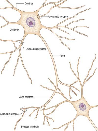
The functional capabilities of the nervous system are a product of its vast population of intercommunicating nerve cells, or neurones, estimated to number on the order of 1010. Neurones encode information, conduct it, sometimes over considerable distances, and then transmit it to other neurones or to non-neural tissues (muscles or glandular cells). Most neurones consist of a central mass of cytoplasm within a limiting cell membrane (the cell body or soma) from which a number of branched processes, termed neurites, extend (Fig. 1.1). One of these, the axon, is usually much longer than the others and normally conducts information away from the cell body. The other processes are termed dendrites, and these typically conduct information toward the soma. The nerve cell membrane is polarized, the inside of the cell being around 70 mV negative with respect to the outside. Information is coded in the form of patterns of transient depolarizations and repolarizations of this membrane potential, known as nerve impulses or action potentials. These are conducted along the axon, which may have collateral branches that permit information to be distributed simultaneously to several targets (Fig. 1.2). Axons possess specialized endings, or axon terminals, that come into close apposition with the membrane of the target cell at synapses, where information passes from one cell to another. Axon terminals may form synaptic contacts with dendrites (axodendritic), cell bodies (axosomatic), other axons (axoaxonic) or non-neural tissue such as muscle cells (neuromuscular junction). Transmission of information to other cells is brought about when action potentials cause the release of specific neurotransmitter substances stored in synaptic vesicles within the presynaptic nerve terminal. Specialized receptors are located on the postsynaptic target cell membrane. The neurotransmitter binds to these and, depending on the nature of the chemical and the receptor, either elicits an excitatory (depolarizing) or inhibitory (hyperpolarizing) response or modulates intracellular second messenger systems.
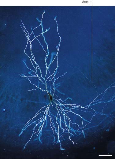
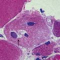
Fig. 1.1 Dark-field illuminated micrograph of a CA3 pyramidal cell in a hippocampal slice culture, intracellularly injected with the dye biocytin. Scale bar 50 µm.
(Courtesy of R. Anne McKinney, McGill University, and Mathias Abegg, Brain Research Institute, University of Zurich.)
The nervous system is customarily divided into two major parts: the central nervous system (CNS) and the peripheral nervous system (PNS). The CNS consists of the brain and spinal cord. The PNS is composed of cranial nerves and spinal nerves together with their ramifications and certain groupings of cell bodies that constitute the peripheral ganglia. Another convention divides the nervous system into somatic and autonomic components. Anatomically, both of these have elements in the CNS and PNS. The autonomic nervous system, which consists of sympathetic and parasympathetic divisions, is made up of neurones concerned primarily with control of the internal environment through innervation of secretory glands and cardiac and smooth muscle. It is considered in detail in Chapter 21. The wall of the gastrointestinal tract contains neurones capable of sustaining local reflex activity independent of the CNS, which are known as the enteric nervous system.
Central Nervous System
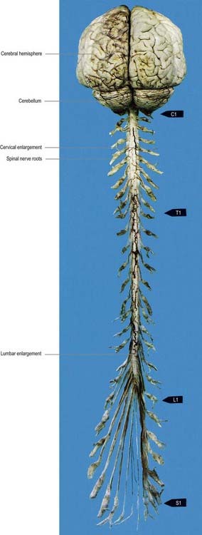
The brain and spinal cord (Fig. 1.3) contain the great majority of neuronal cell bodies in the nervous system. In many parts of the CNS the cell bodies of neurones are grouped together and are more or less segregated from axons. The generic term for such collections of cell bodies is grey matter. Smaller aggregations of neuronal cell bodies, which usually share a common functional role, are termed nuclei. It follows that neuronal dendrites and synaptic interactions are mostly confined to grey matter. Axons tend to be grouped together to form white matter, so called because axons are often ensheathed in myelin, which confers a paler colouration. Axons that pass between similar sources or destinations within the CNS tend to run together in defined pathways or tracts. These often cross the midline (decussate), which means that half of the body is, in many respects, controlled by and sends information to the opposite side of the brain.
Some groups of neurones in the spinal cord and brain stem that subserve similar functions are organized into longitudinal columns. The neurones in these columns may be concentrated into discrete, discontinuous nuclei in some areas, such as the cranial nerve nuclei of the brain stem, or they may form more or less continuous longitudinal bands, as in much of the spinal cord (Fig. 1.4). Efferent neurones constitute three such columns. The somatic motor column contains motor neurones, the axons of which serve muscles derived from head somites. The two other columns are related to specialized features of head morphology. Of these, the branchial motor column innervates muscles derived from the wall of the embryonic pharynx (branchial muscles), and the visceral motor column supplies preganglionic parasympathetic fibres to glands and visceral smooth muscle. There are four longitudinal cell columns related to sensory functions. The general somatic sensory column essentially deals with information from the head. Special somatic sensory neurones are related to the special senses and receive vestibular and auditory input. General visceral sensory neurones deal with information from widespread and varied visceral sensory endings, and special visceral sensory neurones are related to the special sense of taste.
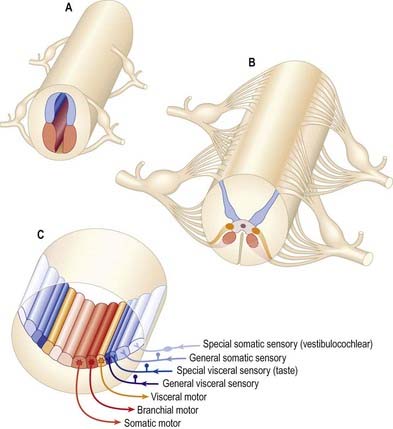
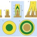
Fig. 1.4 Arrangement of sensory and motor cell columns in the spinal cord and brain stem. A, Organization of the primitive spinal cord with a dorsal sensory column (blue), a ventral column (red) and segmentally arranged dorsal and ventral nerve roots. B, Arrangement of adult spinal cord serving the thorax, with sensory and somatic motor columns colour-coded in the same way as in A, with an additional intermediate (lateral) visceral motor column (orange). C, Arrangement of multiple longitudinal columns in the brain stem, where the motor column is now subdivided into three parts and the sensory column into four. For further information about the embryological aspects of the early nervous system, consult Chapter 3. See also Fig. 10.1.
To sustain the energy required by constant neuronal activity, the CNS has a high metabolic rate and a rich blood supply. The blood–brain barrier controls the neuronal environment and imposes severe restrictions on the types of substances that can pass from the blood stream into nervous tissue.
Spinal Cord
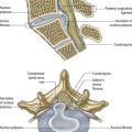
The spinal cord is located within the vertebral column, lying in the upper two-thirds of the vertebral canal (Ch. 8). It is continuous rostrally with the medulla oblongata. For the most part, the spinal cord controls the functions of, and receives afferent input from, the trunk and limbs. Afferent and efferent connections travel in 31 pairs of segmentally arranged spinal nerves. These attach to the cord as dorsal and ventral rootlets that unite to form the spinal nerves proper (Fig. 1.5). The dorsal and ventral roots are functionally distinct. Dorsal roots carry primary afferent nerve fibres from cell bodies located in dorsal root ganglia. Ventral roots carry efferent fibres from cell bodies located in the spinal grey matter.
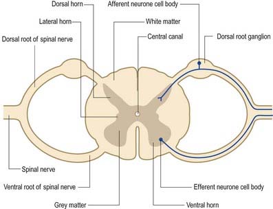
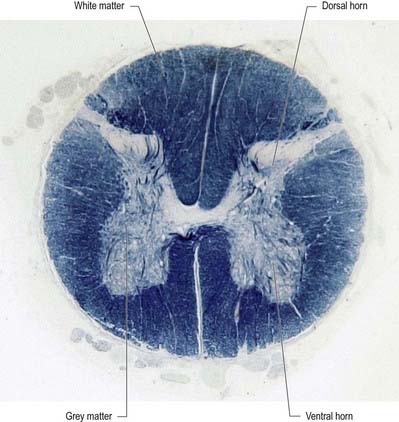
Internally, the spinal cord is differentiated into a central core of grey matter surrounded by white matter. The grey matter is configured in a characteristic H, or butterfly, shape that has projections known as dorsal and ventral horns (Fig. 1.6). In general, neurones situated in the dorsal horn are primarily concerned with sensory functions, and those in the ventral horn are mostly associated with motor activities. At certain levels of the spinal cord a small lateral horn is also present, marking the location of the cell bodies of preganglionic sympathetic neurones. The central canal, which is a vestigial component of the ventricular system, lies at the centre of the spinal grey matter and runs the length of the cord. The white matter of the spinal cord consists of ascending and descending axons that link spinal cord segments to one another and link the spinal cord to the brain.
Brain
The brain (encephalon) lies within the cranium. It receives information from, and controls the activities of, the trunk and limbs, mainly through rich connections with the spinal cord. It possesses 12 pairs of cranial nerves through which it communicates mostly with structures of the head and neck. The brain is divided into major regions on the basis of ontogenetic growth in individuals and phylogenetic principles (Figs. 1.7–1.9; see also Fig. 6.8). Ascending in sequence from the spinal cord, the principal divisions are the rhombencephalon or hindbrain, the mesencephalon or midbrain and the prosencephalon or forebrain.
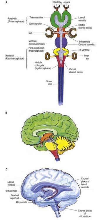
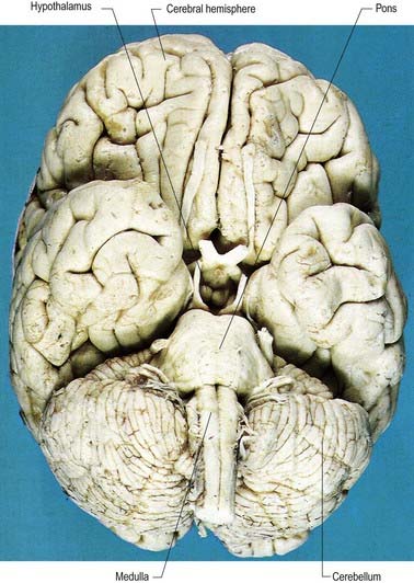
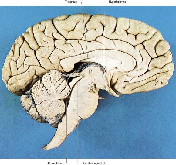
Fig. 1.9 Sagittal section of the brain.
(Photograph by Kevin Fitzpatrick on behalf of GKT School of Medicine, London.)
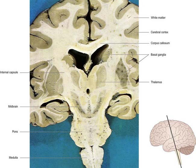
The prosencephalon may be subdivided into the diencephalon and the telencephalon. The diencephalon comprises mostly the thalamus and hypothalamus but also includes the smaller epithalamus and subthalamus. The telencephalon is composed mainly of the two cerebral hemispheres or cerebrum. The diencephalon is almost completely embedded in the cerebrum and is therefore largely hidden. The cerebrum constitutes the major portion of the volume of the human brain. It occupies the anterior and middle cranial fossae and is directly related to the cranial vault. It consists of two cerebral hemispheres. The surface of each hemisphere is convoluted in a complex pattern of ridges (gyri) and furrows (sulci). Internally, each hemisphere has an external layer of grey matter, called the cerebral cortex, beneath which lies a dense mass of white matter (Fig. 1.10). One of the most important components of the cerebral white matter is the internal capsule, which contains nerve fibres that pass to and from the cerebral cortex. Several large nuclei of grey matter, usually referred to as the basal ganglia, are partly embedded in the subcortical white matter. Connections between corresponding areas of the two sides of the brain cross the midline within commissures. By far the largest commissure is the corpus callosum, which links the two cerebral hemispheres.
Overview of Ascending Sensory Pathways
Sensory modalities are conventionally described as either special senses or general senses. The special senses are olfaction, vision, taste, hearing and vestibular function. Afferent information is encoded by highly specialized sense organs and transmitted to the brain in certain cranial nerves (I, II, VII, VIII and IX). The special senses are described in Chapter 12.
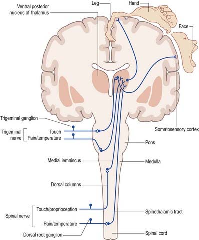
The general senses include touch, pressure, vibration, pain, thermal sensation and proprioception (perception of posture and movement). Stimuli from the external and internal environments activate a diverse range of receptors in the skin, viscera, muscles, tendons and joints. Afferent impulses from the trunk and limbs are conveyed to the spinal cord in spinal nerves, and those from the head are carried to the brain in cranial nerves. The detailed anatomy of the complex pathways by which the various general senses impinge on conscious levels is better understood with reference to certain overall organizational principles. Although undoubtedly oversimplified and subject to many exceptions, this schema is helpful in emphasizing the essential similarities between the ascending sensory systems.
In essence, ascending sensory projections related to the general senses consist of a sequence of three neurones extending from the peripheral receptor to the contralateral cerebral cortex (Fig. 1.11). These are often referred to as primary, secondary and tertiary sensory (afferent) neurones or first-, second- and third-order neurones, respectively. Primary afferents have peripherally located sensory endings, and their cell bodies lie in dorsal root ganglia or the ganglia associated with certain cranial nerves. Their axons enter the CNS through ipsilateral spinal or trigeminal nerves. Within the CNS they terminate ipsilateral to their side of entry, on the cell bodies of second-order neurones. The precise location of this termination depends on the modality.
Overview of Descending Motor Pathways
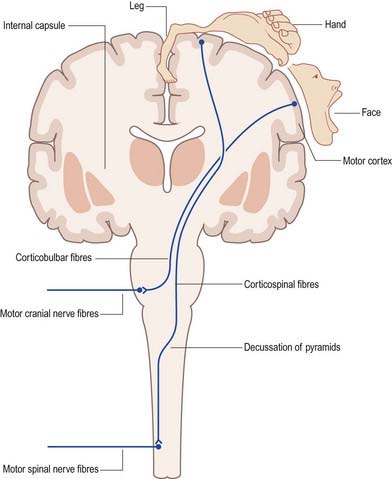
The concept of upper and lower motor neurones is fundamental to the clinical description of the effects of lesions of the motor system. The term ‘lower motor neurones’ refers to the alpha motor neurones that innervate the extrafusal muscle fibres of skeletal muscle. The term ‘upper motor neurones’ in theory refers collectively to all the descending pathways that impinge on the activity of lower motor neurones. In common parlance, however, the term is often equated with the corticospinal (pyramidal) tract (Fig. 1.12). This pathway originates from widespread regions of the cerebral cortex, including the primary motor cortex of the frontal lobe, where the opposite half of the body is represented in a detailed somatotopic fashion. Corticofugal fibres descend through the internal capsule and pass into the brain stem, where some of them (designated corticobulbar fibres) terminate. Corticobulbar fibres control the activity of brain stem neurones, including motor neurones within the cranial nerve nuclei. Corticospinal fibres descend through the brain stem. The majority of them cross to the contralateral side in the pyramidal decussation of the medulla and continue as the lateral corticospinal tract of the spinal cord. This terminates in association with interneurones and motor neurones of the spinal grey matter. The principal function of the corticospinal and corticobulbar tracts is the control of fine, fractionated movements, particularly of those parts of the body where delicate muscular control is required. These tracts are particularly important in speech (corticobulbar tract) and movement of the hand (corticospinal tract).
The terms ‘upper motor neurone lesion’ and ‘lower motor neurone lesion’ are used clinically to distinguish, for example, between the effects of a stroke in the internal capsule (a typical upper motor neurone lesion) and those of motor neurone disease (a typical lower motor neurone lesion). These produce very different signs and symptoms (summarized below), which are indicative of the anatomical site of the lesion.
Two other major systems that contribute to the control of movement are the basal ganglia and the cerebellum. The basal ganglia are a group of large subcortical nuclei, the major components of which are the caudate nucleus, putamen and globus pallidus (see Fig. 1.10; Ch. 14). These structures have important connections with the cerebral cortex, certain diencephalic nuclei of the thalamus and subthalamus and the brain stem. They appear to be involved in the selection of appropriate movements and the suppression of inappropriate ones. Disorders of the basal ganglia cause either too little movement (akinesia) or abnormal involuntary movements (dyskinesia), as well as tremor and abnormalities of muscle tone. The basal ganglia are sometimes described as being part of the so-called extrapyramidal (motor) system. This term is used to distinguish between the effects of basal ganglia disease and the effects of damage to the pyramidal (corticospinal) system. However, the progressive elucidation of basal ganglia anatomy and the pathophysiology of motor disorders has revealed the close functional interrelationship between the two ‘systems’ and has rendered the terms that distinguish them largely obsolete (Brodal 1981). The cerebellum has rich connections with the brain stem, particularly the reticular and vestibular nuclei, and with the thalamus. It is concerned with the coordination of movement. Among the clinical signs of cerebellar disorders are ataxia, hypotonia and the so-called intention tremor.
Peripheral Nervous System
Spinal Nerves
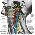
Spinal nerves are the means by which the CNS receives information from, and controls the activities of, the trunk and limbs. Spinal nerves are considered in detail elsewhere. In brief, there are 31 pairs of spinal nerves (8 cervical, 12 thoracic, 5 lumbar, 5 sacral, 1 coccygeal) that contain a mixture of sensory and motor fibres. They originate from the spinal cord as continuous lines of dorsal and ventral nerve rootlets. Adjacent groups of rootlets fuse to form dorsal and ventral roots, which then merge to form the spinal nerves proper. The dorsal roots of spinal nerves contain afferent nerve fibres from cell bodies located in dorsal root ganglia. These cells give off both centrally and peripherally directed processes and do not have synapses on their cell bodies. The ventral roots of spinal nerves contain efferent fibres from cell bodies located in the spinal grey matter. They include motor neurones innervating skeletal muscle and preganglionic autonomic neurones.
Cranial Nerves
Cranial nerves are the means by which the brain receives information from, and controls the activities of, the head and neck and, to a lesser extent, the thoracic and abdominal viscera. The cranial nerves’ component fibres, their route of exit from the cranial cavity, their subsequent peripheral course and their distribution and functions are considered in detail elsewhere. Their origins, destinations and connections within the CNS are considered in this section.
Briefly, there are 12 pairs of cranial nerves. They are individually named and numbered (I to XII) in a rostrocaudal sequence (Table 1.1). Unlike spinal nerves, only some are mixed in function and carry both sensory and motor fibres. Others are purely sensory or purely motor. The first cranial nerve (I; olfactory) has an ancient lineage and is derived from the forerunner of the cerebral hemisphere. It retains this unique position through the connections of the olfactory bulb, and it is the only sensory cranial nerve that projects directly to the cerebral cortex rather than via the thalamus, as do all other sensory modalities. The areas of cerebral cortex involved have a primitive cellular organization and are an integral part of the limbic system, which is concerned with the emotional aspects of behaviour. The second cranial nerve (II; optic) consists of the axons of second-order visual neurones and terminates in the thalamus. The other 10 pairs of cranial nerves attach to the brain stem. Most of the component fibres originate from or terminate in named cranial nerve nuclei.
| Number | Name | Function |
|---|---|---|
| I | Olfactory | Olfaction |
| II | Optic | Vision |
| III | Oculomotor |
a
Brodal, 1981Brodal A. Neurological Anatomy in Relation to Clinical Medicine, third ed. 1981. Oxford. Oxford University Press.
Crossman A.R., Neary D. Neuroanatomy: An Illustrated Colour Text, second ed. Churchill Livingstone: Edinburgh; 2000.
England M.A., Wakely J. A Colour Atlas of the Brain and Spinal Cord. Wolfe Publishing Ltd: London; 1991.
Haines D.E. Neuroanatomy: An Atlas of Structures, Sections and Systems, fifth ed. Lippincott Williams & Wilkins: Philadelphia; 2000.