Borderline Ovarian Tumor

Published on 10/03/2015 by admin

Filed under Obstetrics & Gynecology

Last modified 22/04/2025

Print this page

rate 1 star rate 2 star rate 3 star rate 4 star rate 5 star
Your rating: none, Average: 0 (0 votes)

This article have been viewed 1687 times

Borderline Ovarian Tumor

Synonyms/Description

Tumor of low malignant potential (LMP tumor)

Etiology

Ten to fifteen percent of epithelial ovarian tumors are considered borderline malignancies. They tend to occur in women in their forties and fifties, younger than those with frankly invasive tumors, and they are not thought to be related to hereditary breast/ovarian cancer syndromes. These tumors are stage I at diagnosis in more than 90% of patients, have infrequent recurrence, and an excellent prognosis. The survival rates for stage I and stage III are close to 100% and 80% to 90%, respectively. The survival is good even in patients who have developed peritoneal spread. Just more than half are serous, with the rest being mucinous and occasional mixed intestinal cell or endometrioid types. The mucinous tumors tend to be unilateral and are most often of the intestinal type, with a minority being of the endocervical type. Serous tumors have a higher incidence of bilaterality.

Ultrasound Findings

The typical ultrasound appearance of a borderline ovarian cancer is a complex cystic mass with septations, nodularity/papillations, and irregular walls. Flow is typically identified in the solid areas, although not so much flow nor so many irregular vessels as are usually seen in invasive cancers. This difference is neither sufficient nor reliable enough to differentiate invasive from borderline tumors accurately sonographically.
In a study of 113 borderline ovarian tumors, the mucinous tumors of the intestinal type tended to be the largest compared with those of the cervical type or serous lesions. The serous and the mucinous endocervical type of tumors also tended to have a higher number of papillary excrescences and a lower percentage of multilocular lesions when compared with the intestinal type. Solid tumors were found only among the serous tumors.

Differential Diagnosis

The sonographic appearance of borderline tumors is often similar to that of invasive cancers. Other diagnostic possibilities include a decidualized endometrioma in a pregnant woman (see Endometriosis) or a cystadenofibroma. Typically the cystadenofibromas and benign cystadenomas have smaller areas of nodularity with little if any blood flow within the nodule or septae.

Clinical Aspects and Recommendations

The vast majority of mucinous borderline tumors are stage I, with typically benign behavior. On the other hand, the less common endocervical type of mucinous tumor has a worse prognosis, presenting with invasive implants more frequently and recurring more often than the intestinal type. Serous borderline tumors are stage I at presentation in 65% to 70% of cases with a 5-year survival of 95% when there are noninvasive implants and 66% in patients with invasive implants.
The prognosis of patients with borderline ovarian tumors is largely based on the presence of invasive peritoneal implants or residual disease after surgery.
Treatment is surgical removal as well as staging. In patients who have not completed child bearing, the uterus and contralateral ovary may be preserved.
Increasingly in stage I disease, ovarian cystectomy only and close observation are being advocated for those who have not completed child bearing.

Figures

image

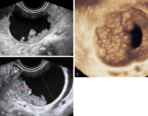
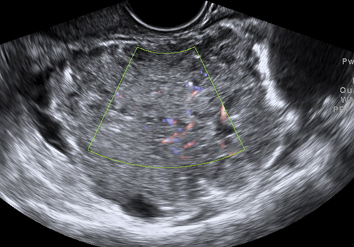
Figure B2-1 A papillary serous cystadenoma with irregular nodularity. A, The lateral side of the mass. B, The medial side with thicker nodularity. Note the modest blood flow in the solid areas, less than would be expected in a more aggressive tumor. C, A 3-D image of the surface texture of the inner aspect of the tumor.

image

Figure B2-2 A mucinous borderline tumor. Note the abundant solid portions and unilocular and large lesion.


image

Figure B2-3 A and B, Borderline serous papillary tumor. Note the papillations and color Doppler, indicating flow in the solid areas. C, The papillations using 3-D surface imaging.


image

Figure B2-4 Bilateral serous borderline tumors. A and B, The left ovarian mass with the small lesion containing a single vascular nodule. C, The right ovarian mass is larger, with low-level echoes and a large vascular nodule. D, A 3-D volume illustrating the surface of the nodularity.


image

Figure B2-5 Color flow is seen using Doppler in the septation of this borderline tumor.

image

Figure B2-6 The unusually solid appearance of a borderline tumor, mimicking an invasive cancer.


Suggested Reading

Behtash N., Modares M., Abolhasani M., Ghaemmaghami F., Mousavi M., Yarandi F., Hanjani P. Borderline ovarian tumours: clinical analysis of 38 cases. J Obstet Gynaecol.. 2004;24:157–160.

Fruscella E., Testa A.C., Ferrandina G., De Smet F., Van Holsbeke C., Scambia G., Zannoni F.G., Ludovisi M. Ultrasound features of different histopathological subtypes of borderline ovarian tumors. Ultrasound Obstet Gynecol. 2005;26:644–650.

Morice P., Uzan C., Fauvet R., Gouy S., Duvillard P., Darai E. Borderline ovarian tumour: pathological diagnostic dilemma and risk factors for invasive or lethal recurrence. Lancet Oncol.. 2012;13:103–115.

Tropé C.G., Kaern J., Davidson B. Borderline ovarian tumours. Best Pract Res Clin Obstet Gynaecol. 2012;26:325–336.