Ovarian Cancer

Published on 10/03/2015 by admin

Filed under Obstetrics & Gynecology

Last modified 22/04/2025

Print this page

rate 1 star rate 2 star rate 3 star rate 4 star rate 5 star
Your rating: none, Average: 0 (0 votes)

This article have been viewed 1398 times

Chapter 39 Ovarian Cancer

Ovarian cancer is the fifth most common cancer among females in the United States, accounting for one fourth of all gynecologic cancers. It is the leading cause of death from gynecologic cancer because it is difficult to detect before it disseminates. In 2007, 22,430 new cases and more than 15,280 deaths are expected from this disease. Most women with ovarian cancer are in the fifth or sixth decade of life.

image Etiology and Epidemiology

The cause of ovarian cancer is unknown. The patient characteristics found to be associated with an increased risk for epithelial ovarian cancer include white race, late age at menopause, family history of cancer of the ovary, breast, or bowel, and prolonged intervals of ovulation uninterrupted by pregnancy. There is an increased prevalence of ovarian cancer in nulliparous women and those who have been infertile.

The incidence of ovarian cancer varies in different geographic locations. Western countries, including the United States, have rates that are 3 to 7 times greater than those in Japan. Second-generation Japanese immigrants to the United States have an incidence of ovarian cancer similar to that of American women. White Americans experience ovarian cancer about 1.5 times more frequently than do black Americans.

About 10% of epithelial ovarian cancers occur in women with a hereditary predisposition. In women with hereditary cancers, two or more first-degree relatives on either the paternal or maternal side typically have had breast or ovarian cancer. The pattern of inheritance is autosomal dominant. Breast cancers generally occur in young premenopausal women, whereas ovarian cancers have a median age of about 50 years. The breast-ovarian cancer syndrome is due to germline mutations of BRCA1, which is located on chromosome 17, and BRCA2, which is located on chromosome 13. The Lynch II syndrome, nonpolyposis colorectal cancer syndrome, is associated with mutations in the mismatch repair genes. Adenocarcinomas of the ovary, breast, colon, stomach, pancreas, and endometrium are seen in the families of these individuals.

The use of oral contraceptives has been found to protect against ovarian cancer, possibly because of suppression of ovulation. It has been postulated that incessant ovulation may predispose to malignant transformation in the ovary.

Patients with a known germline mutation (e.g., BRCA1 and BRCA2 mutations) may be offered prophylactic salpingo-oophorectomy once childbearing has been completed, and this operation is highly protective for ovarian and fallopian tube carcinomas. Indeed, the risk for subsequent breast cancer is also significantly reduced in these women. There is still a small risk for peritoneal carcinoma after prophylactic salpingo-oophorectomy.

Some case-control studies have suggested that the use of postmenopausal estrogen replacement therapy may increase the risk for ovarian cancer, but these data are controversial.

It has also been postulated that a causative agent could enter the peritoneal cavity through the lower genital tract. For example, the perineal use of asbestos-contaminated talc has been linked to the development of epithelial ovarian cancer. This possibility remains controversial, although tubal ligation and hysterectomy are both associated with a decreased risk for the disease.

image Preoperative Evaluation

The diagnosis of ovarian cancer requires a laparotomy or laparoscopy. Routine preoperative hematologic and biochemical studies should be obtained, as should a chest radiograph. A pelvic and abdominal computed tomography scan will exclude liver metastases, but it is not mandatory.

A Papanicolaou smear should be obtained to evaluate the cervix, but this test is of limited value in detecting ovarian cancer. Endometrial biopsy and endocervical curettage are necessary in patients with abnormal vaginal bleeding because concurrent primary tumors occasionally occur in the ovary and endometrium. In the presence of a pelvic mass, it is preferable not to perform abdominal paracentesis for cytologic evaluation of ascitic fluid, unless neoadjuvant chemotherapy is planned, because seeding of the abdominal wall may occur.

An abdominal radiograph may be useful in a younger patient to locate calcifications associated with a benign cystic teratoma (dermoid cyst), which is the most common neoplasm in patients younger than 25 years of age. In patients with occult blood in the stool or significant intestinal symptoms, a barium enema or lower gastrointestinal endoscopy should be obtained to rule out a primary colonic cancer with ovarian metastasis.

Similarly, an upper gastrointestinal endoscopy is important if there are significant gastric symptoms. Breast cancer may also metastasize to the ovaries, so bilateral mammograms should be obtained if there are any suspicious breast masses.

Pelvic ultrasonography, particularly transvaginal ultrasonography with or without color Doppler studies, may be useful for smaller (<8 cm) masses in premenopausal women. Masses that are predominantly solid or multilocular have a high probability of being neoplastic, whereas unilocular cystic masses are generally functional cysts. In postmenopausal women, ultrasonography may also be useful because small, unilocular cysts (<5 cm) that are stable are generally benign.

Several tumor markers have been investigated, but none has been consistently reliable. The tumor-associated antigen CA 125 is elevated in only about 50% of women with stage I ovarian cancer. When this assay is elevated, it is useful for monitoring the clinical course of the disease.

image Staging

The standard staging system for ovarian cancer is presented in Table 39-1. Ovarian cancer is surgically staged according to the International Federation of Gynecology and Obstetrics (FIGO) staging system.

TABLE 39-1 INTERNATIONAL FEDERATION OF GYNECOLOGY AND OBSTETRICS (FIGO) STAGING FOR PRIMARY CARCINOMA OF THE OVARY

Stage I Growth limited to the ovaries
Stage Ia Growth limited to one ovary; no ascites. No tumor on the external surface; capsule intact
Stage Ib Growth limited to both ovaries; no ascites. No tumor on the external surfaces; capsules intact
Stage Ic Tumor either stage Ia or Ib but with tumor on the surface of one or both ovaries or with capsule ruptured or with ascites present containing malignant cells or with positive peritoneal washings
Stage II Growth involving one or both ovaries with pelvic extension
Stage IIa Extension or metastases, or both, to the uterus or tubes, or both
Stage IIb Extension to other pelvic tissues
Stage IIc Tumor either stage IIa or IIb but with tumor on the surface of one or both ovaries or with capsule or capsules ruptured or with ascites present containing malignant cells or with positive peritoneal washings
Stage III Tumor involving one or both ovaries with peritoneal implants outside the pelvis or positive retroperitoneal or inguinal nodes, or both. Superficial liver metastasis equals stage III. Tumor is limited to the true pelvis, but with histologically proven malignant extension to small bowel or omentum
Stage IIIa Tumor grossly limited to the true pelvis with negative nodes but with histologically confirmed microscopic seeding of abdominal peritoneal surfaces
Stage IIIb Tumor of one or both ovaries with histologically confirmed implants of abdominal peritoneal surfaces, none exceeding 2 cm in diameter. Nodes negative for disease
Stage IIIc Abdominal implants >2 cm in diameter or positive retroperitoneal or inguinal nodes, or both
Stage IV Growth involving one or both ovaries with distant metastasis. If pleural effusion is present, there must be positive cytologic test results to allot a case to stage IV. Parenchymal liver metastasis equals stage IV.

Even though all microscopic disease may appear to be confined to the ovaries at the time of laparotomy, microscopic spread may have already occurred; thus, patients must undergo a thorough surgical staging. Procedures necessary to stage ovarian cancer are shown in Box 39-1.

image Classification

The histologic classification of ovarian neoplasms is listed in Table 39-2. These lesions fall into four categories according to their tissue of origin. Most ovarian neoplasms (80% to 85%) are derived from coelomic epithelium and are called epithelial carcinomas. Less common tumors are derived from primitive germ cells, specialized gonadal stroma, or nonspecific mesenchyme. In addition, the ovary can be the site of metastatic carcinomas, most often from the gastrointestinal tract or the breast.

TABLE 39-2 HISTOGENETIC CLASSIFICATION OF PRIMARY OVARIAN NEOPLASMS

Derivation Type of Tumor
Coelomic epithelial origin (80%-85%)

Germ cell origin (10%-15%) Specialized gonadal-stromal origin (3%-5%) Nonspecific mesenchymal origin (fewer than 1%)

Combined germ cell and specialized gonadal-stromal elements.

Data from Hart WR, Morrow CP: The ovaries. In Romney SL, Gray MJ, Little AO, et al (eds): Gynecology and Obstetrics: The Health Care of Women, 2nd ed. New York, McGraw-Hill, 1981.

image Epithelial Ovarian Carcinomas

PATHOLOGIC FEATURES

The main histologic subtypes of epithelial carcinomas are serous (about 55%), mucinous (about 20%), endometrioid (about 15%), and clear cell (about 5%). Malignant Brenner tumors and undifferentiated carcinomas are uncommon.

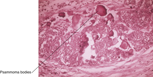
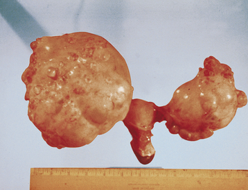
Serous tumors resemble fallopian tube epithelium histologically (Figure 39-1). About 30% of patients with stage I and stage IIa disease have bilateral involvement. On gross examination, serous carcinomas have an irregular and multilocular appearance (Figure 39-2).

Mucinous tumors histologically resemble endocervical epithelium and are often large, measuring 20 cm or more in diameter. They are bilateral in 10% to 20% of patients.

Endometrioid tumors closely resemble carcinomas of the endometrium and arise in association with primary endometrial cancer in about 20% of patients. In early-stage disease, they are bilateral in about 10% of cases. About 10% of endometrioid ovarian carcinomas are associated with endometriosis, although malignant transformation of endometriosis occurs in fewer than 1% of patients.

Clear cell carcinomas of the ovary are uncommon. In about 25% of cases, they occur in association with endometriosis.

The Brenner tumor represents only 2% to 3% of all ovarian neoplasms, and fewer than 2% of these tumors are malignant. About 10% of Brenner tumors occur in conjunction with a mucinous cystadenoma or dermoid cyst in the same or opposite ovary.

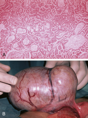
Tumors of low malignant potential or borderline histologic appearance exist for each histologic type. About 5% to 10% of malignant serous tumors are borderline (Figure 39-3), whereas 20% of malignant mucinous tumors fall into this category. The endometrioid, clear cell, and Brenner tumors are only rarely borderline.

MANAGEMENT OF EPITHELIAL OVARIAN CANCER

The initial approach to all patients with ovarian cancer is surgical exploration of the abdomen and pelvis.

Early-Stage Disease

Definitive diagnosis requires an intraoperative frozen section. In patients with no gross evidence of disease beyond the ovary, the standard operation is total abdominal hysterectomy, bilateral salpingo-oophorectomy, infracolic omentectomy, and thorough surgical staging, as shown in Box 39-1. Patients who wish to preserve fertility may have a unilateral salpingo-oophorectomy. In patients with grade 1 or 2 tumors confined to one or both ovaries after surgical staging, no further treatment is necessary. Patients with poorly differentiated (grade 3) tumors are subsequently treated with systemic chemotherapy.

Advanced-Stage Disease

In patients with advanced disease, cytoreductive surgery (“debulking”) is required. The objectives are to remove the primary tumor and all of the metastases, if possible. If all macroscopic disease cannot be removed, an attempt should be made to reduce individual tumor nodules to 1 cm or less in diameter. Patients in whom this goal is achieved are said to have had “optimal” cytoreduction, which can be achieved in about 70% of patients. In addition to a total or subtotal abdominal hysterectomy, bilateral salpingo-oophorectomy, omentectomy, and resection of peritoneal metastases, optimal cytoreduction may necessitate bowel resection; therefore, all patients having surgery for suspected ovarian cancer should have a bowel preparation preoperatively.

In retrospective studies, patients whose individual residual tumor nodules are 1 cm in diameter or less before the commencement of chemotherapy have been shown to have longer median survivals and more complete responses to therapy. The longest survival is seen in patients in whom all visible tumor has been removed before treatment.

In patients who are medically unfit or have a poor performance status, usually because of a large pleural effusion and massive ascites, it may be prudent to give two or three cycles of neoadjuvant chemotherapy before undertaking radical surgery. If the disease does not respond to chemotherapy, as evidenced by the failure to resolve the malignant effusions, the patient should be offered palliative care only. Usually, the effusions resolve completely, and an “interval” cytoreductive operation can be safely undertaken.

Following primary cytoreductive surgery, combination chemotherapy is given, most commonly intravenous carboplatin and paclitaxel, or intraperitoneal cisplatin and paclitaxel. Intraperitoneal treatment is only useful for patients with minimal residual disease. Single-agent therapy with paclitaxel, or carboplatin, is occasionally used for frail or elderly patients. During chemotherapy, the patient’s response is monitored with serial CA 125 levels. If the values rise or plateau within 6 months, it is advisable to change to second-line drugs, such as liposomal-encapsulated doxorubicin, topotecan, etoposide, gemcitabine, or experimental chemotherapeutic agents. If the progression-free interval has been longer than 6 to 12 months, the patient may respond to further paclitaxel or carboplatin chemotherapy. Response to second-line chemotherapy is in the range of 20% to 50%, but patients are not considered to be curable after their initial relapse. Secondary cytoreduction may be appropriate if the disease-free interval is 24 months or longer.

It is unclear whether patients with “metastatic” borderline tumors benefit from chemotherapy.

image Germ Cell Tumors

Germ cell tumors of the ovary account for only about 2% to 3% of all ovarian malignancies. They occur predominantly in young patients and frequently produce either human chorionic gonadotropin (hCG) or alpha-fetoprotein (AFP), which serve as tumor markers. The most common germ cell tumors are the dysgerminoma and immature teratoma. Endodermal sinus tumors, embryonal tumors, and nongestational choriocarcinomas are less common. Mixed germ cell tumors are not uncommon.

DYSGERMINOMAS

Dysgerminomas occur predominantly in children and young women. About 10% are bilateral. These tumors, of varying malignant virulence, are occasionally seen in patients with gonadal dysgenesis or the testicular feminization syndrome. In such patients, the dysgerminoma may arise in a gonadoblastoma. In about two thirds of patients, the disease is confined to the ovaries at the time of diagnosis. About 10% of dysgerminomas are associated with other germ cell malignancies. Pure dysgerminomas do not produce the tumor markers hCG and AFP but commonly produce lactate dehydrogenase.

image Specialized Gonadal-Stromal Tumors

A group of relatively uncommon tumors is derived from the specialized ovarian stroma. As such, they are often endocrinologically functional, many of them being capable of synthesizing gonadal or adrenal steroid hormones. Because the ovarian stroma has sexual bipotentiality, hormones that are secreted can be either female or male. Estrogen and progesterone are typically associated with granulosa-theca cell tumors, whereas testosterone and other androgens may be secreted by many Sertoli-Leydig cell tumors. Rarely, lipid cell tumors, which are usually virilizing, produce adrenal corticoids and a clinical cushingoid syndrome.

image Fallopian Tube Carcinoma

Primary carcinoma of the fallopian tube accounts for only 0.1% to 0.5% of gynecologic cancers and is diagnostically confused with ovarian carcinoma. The incidence of fallopian tube carcinoma may be higher than previously suspected because some are misclassified as primary ovarian carcinomas. Most are adenocarcinomas, but sarcomas and mixed tumors can occur. There is no official staging system for fallopian tube carcinoma, but in general they are staged like ovarian cancer because the mode of dissemination is similar. Bilateral carcinomas are seen in 10% to 20% of patients.