52
Otitis externa
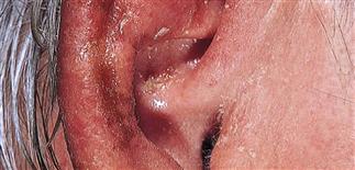
The ear shows marked near confluence of erythema of varying intensity. There is scale crust and some weeping in the bowl of the ear. A culture is warranted.
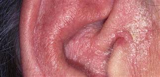
Severe external otitis of the entire ear. The ear is swollen, tender, and crusted. A bacterial source should be considered, and awareness of potential contact allergens is prudent.
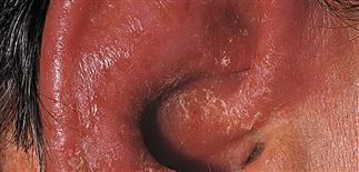
Mild inflammation with scaling and a faint pink erythema anterior to the ear, suggesting psoriasis or seborrheic dermatitis. This will usually resolve with a mild topical steroid solution.
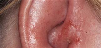
Intense inflammation of the pinna with swelling and exudate. Pseudomonas was cultured. Treatment was with levofloxacin.
DESCRIPTION
Otitis externa is an inflammation of the external auditory canal, usually with secondary infection.
HISTORY
• A mild, self-limited form known as swimmer’s ear is common in children. • Symptoms range from itch and irritation to severe pain. • Mechanical cleansing of the external canal and medicaments used to relieve symptoms can mask or exacerbate the condition. • Ear wax forms a water-resistant barrier for the thin skin that lines the canal, and inhibits bacterial growth by maintaining a low pH environment. When this protective barrier is disrupted, bacterial overgrowth can occur. • The usual pathogen is Pseudomonas, but mixed infections with Staphylococcus and Pseudomonas species are common. • Swimmer’s ear typically caused by Pseudomonas species, Enterobacteriaceae, or Proteus species. Acute infection usually due to Staphylococcus aureus. • Secondary infection with Candida species may occur but is uncommon. • A rare and severe form of otitis, referred to as malignant external otitis, may develop in patients who have diabetes or who have had ear surgery.
PHYSICAL FINDINGS
• External auditory canal is inflamed with erythema, edema, and dull pain. • Keratin and inflammatory cell debris accumulate within canal. • Most cases do not progress. With progression, pinna becomes red, hot, and edematous, and may develop purulent drainage. • Cellulitis involves entire pinna and often extends to preauricular skin. Pain becomes sharp and constant. • External auditory canal should be cultured.
TREATMENT
• Treatment involves reestablishing the natural protective barrier. • Cellular debris is flushed from the external canal with gentle irrigation. • An acetic acid solution (VoSol otic solution or VoSol HC otic solution) helps lower pH and inhibits bacterial and fungal growth. • Ofloxacin otic solution 0.3% (Floxin otic solution twice daily) or ciprofloxacin and hydrocortisone (Cipro HC otic) are instilled twice a day. For acute disease, culture to determine organism and sensitivity. • Topical steroids, wet dressings using an astringent such as Domeboro, or Bluboro wet dressings applied for 30 min three times daily, and oral antibiotics such as ciprofloxacin (Cipro) are valuable when cellulitis involves pinna. • Avoid the more common contact allergens (such as topical bacitracin, neomycin, hydrocortisone) and minimize other topical products. • Discourage vigorous scratching, rubbing, or mechanical debridement of external auditory canal. • Malignant external otitis requires hospitalization, administration of i.v. antibiotics, and debridement, as well as consideration of a computerized or magnetic resonance imaging scan to evaluate possible osteomyelitis. • Otolaryngology consultation recommended for malignant external otitis.


