64. Osler-Weber-Rendu Syndrome
Definition
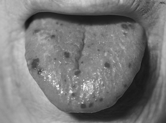
Osler-Weber-Rendu syndrome (OWRS) is an autosomal dominant, inherited disorder characterized by telangiectases, recurrent epistaxis, and first-degree relative(s) positive for the disorder. Telangiectases are lesions produced by telangiectasia. They may present as a coarse or fine, red line or may appear as a punctum with radiating arms (spider-like appearance). Osler-Weber-Rendu syndrome is also known as hereditary hemorrhagic telangiectasia (HHT).
Incidence
The incidence of OWRS is 1:100,000 to 2:100,000. It is reported in all racial groups and almost all ethnic groups without predilection for either gender.
Curaçao Criteria for Osler-Weber-Rendu Syndrome
• Epistaxis: Spontaneous and recurrent
• Family history: First-degree relative positive for hereditary hemorrhagic telangiectasia
• Telangiectases: Multiple at various characteristic sites (lips, oral cavity, fingers, nose)
• Visceral lesions: Gastrointestinal telangiectasia, pulmonary AVM, hepatic AVM, central AVM, or spinal AVM
AVM, Arteriovenous malformation.
Etiology
There are four types of HHT: (1) HHT type 1, (2) HHT type 2, (3) JPHT, and (4) HHT type 3. Genetic loci have been identified for three types of the disease. HHT type 1 results from defects in the long arm of chromosome 9 (9q33-34). HHT type 2 is derived from defects on the long arm of chromosome 12 (12q13). JPHT appears to result from mutations on the long arm of chromosome 5 (5q31.1-32). HHT type 3 appears to follow pathways similar to the three other types, but a specific gene locus has not been identified.
 |
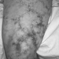
| Osler-Weber-Rendu Syndrome. Note the telangiectatic lesions. |
Signs and Symptoms
• Anemia
• Arteriovenous malformations (AVM) at multiple sites, notably the brain and lungs
• Clubbing
• Continuous thoracic bruit
• Cyanosis
• Dyspnea on exertion
• Easy fatigability
• Epistaxis
• Hemoptysis
• Hepatomegaly
• Jaundice
• Migraine
• Pallor
• Paraparesis
• Polycythemia
• Seizure
• Tachypnea
• Telangiectases of skin, mucous membranes, liver, brain, and spleen
Medical Management
Medical care for the patient with HHT focuses on decreasing hemorrhage, minimizing blood loss, and minimizing sequelae from chronic blood loss. The most common source of blood loss is recurrent epistaxis. The epistaxis may be treated via several methods, including humidification, nasal packing, estrogen therapy, aminocaproic acid, electrocautery, laser ablation, and septal dermoplasty. Blood transfusion may be necessary to correct chronic blood losses.
Hereditary Hemorrhagic Telangiectasia: Medical Interventions by Specific Affected Area
• Central nervous system AVM: Embolization/embolotherapy, stereotactic radiosurgery
• Epistaxis: Humidification, packing, estrogen therapy, aminocaproic acid, electrocautery, ND:YAG laser ablation, transfusion
• GI bleeding: Estrogen/progesterone therapy, aminocaproic acid, endoscopic photoablation, electrocautery, transfusion
• Pulmonary AVM: Embolization via transluminal deployment of a balloon or coil
• Skin lesion: Laser ablation, hypertonic saline sclerotherapy, topical medications
AVM, Arteriovenous malformation; ND:YAG, neodmium–yttrium–garnet.
Surgical interventions may be indicated for several body areas. Epistaxis may be severe enough to warrant septal dermoplasty. Arteriovenous malformations (AVMs) are somewhat weakened outcroppings within the arterial-venous continuum and are prone to enlargement and rupture over time. AVMs are characteristic of OWRS and are particularly disconcerting when they are found in the lungs and/or brain, where rupture can lead to rapid demise. Surgical resection may be undertaken to avert/avoid rupture of an AVM and any potential associated untoward sequelae.
Gastrointestinal bleeding may occur episodically and may be a contributing source of chronic blood loss. Surgical intervention for gastrointestinal bleeding is indicated if the episode is believed to be a massive hemorrhagic event.
Complications
• Abdominal angina
• Brain abscess
• Encephalopathy
• Epidural hematoma
• Hemoptysis
• High-output heart failure
• Intraocular hemorrhage
• Ischemic stroke
• Mesenteric arterial “steal”
• Paradoxical emboli
• Paraparesis
• Portal hypertension
• Pseudocirrhosis
• Severe right-to-left pulmonary shunt
• Subarachnoid hemorrhage
• Transient ischemic attack
Anesthesia Implications
Telangiectatic lesions may occur throughout the body, including mucous membranes. While carrying out endotracheal intubation via direct laryngoscopy, the anesthetist must be aware of the potential for rupturing telangiectases, which may produce a large volume of blood loss. The bleeding can quickly obscure and obliterate the anesthetist’s view of the airway anatomy. For this reason, direct laryngoscopy must be relatively gentle but at the same time accomplished quickly to avoid aspiration of blood if a telangiectatic lesion does rupture. Placement of a laryngeal mask airway should be avoided because of the potential for rupture and hemorrhage from telangiectatic lesions.
Because of the fragility of AVMs, the anesthetist must strive to avoid extreme swings in this patient’s blood pressure throughout the duration of the anesthetic. The anesthetist should also strive to reduce the inspiratory pressure during mechanical ventilation. This may be achieved by using the pressure-controlled ventilation and setting a relatively low peak inspiratory pressure. In addition, the expiratory phase should be prolonged, on the order of 1:2.5 or 1:3, to prevent air trapping that may contribute to rupture of pulmonary AVMs. The tidal volumes of positive pressure ventilations should be reduced when the number of breaths per minute increases to achieve the “calculated” minute ventilation the patient needs. These interventions will reduce the possible trauma to any pulmonary AVM that may be present. Appearance of blood in the endotracheal tube may indicate rupture of a pulmonary AVM.
The anesthetist must also be alert for signs and symptoms of a subarachnoid hemorrhage (particularly during non-neurovascular surgery), which closely follow signs and symptoms of increased intracranial pressure, such as widening pulse pressure, bradycardia, and dilated pupils. Epidural hematoma formation is also a significant risk during surgery. The anesthetist should watch closely for the development of an epidural hematoma in patients with newly demonstrated lower extremity weakness or paralysis.
Cirrhosis in the patient with OWRS is indicative of liver function impairment. Diminution of liver function may alter the metabolism of several anesthesia-related drugs, such as sodium thiopental, midazolam, opiate analgesics, and muscle relaxants. In such events, doses of individual medications that depend heavily on the liver for metabolism/biotransformation may need to be reduced. Severe instances of arteriovenous shunting within the liver may result in the high-output form of liver failure.