Chapter 47 Orthopedic Emergencies
2 How are most cases of osteomyelitis acquired? Direct inoculation? Hematogenous spread? Contiguous spread?
Kaplan SL: Osteomyelitis in children. Infect Dis Clin North Am 19:787–797, 2005.
4 What imaging studies are indicated when osteomyelitis is suspected?
 Plain radiography is a reasonable initial imaging choice in the emergency department (ED) evaluation of a patient with suspected osteomyelitis. This may rule out fracture, tumor, or other concerns. Its usefulness is limited, however, because bony changes (lytic lesions, periosteal elevation, and periosteal new bone formation) may not appear until 10–20 days after symptoms begin. Changes to adjacent soft tissues (deep soft tissue swelling and loss of normal tissue planes) may occur much earlier, as early as several days after symptoms begin.

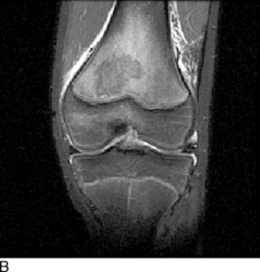
 Magnetic resonance imaging (MRI) (Fig. 47-1) appears to be the imaging study of choice for evaluating the patient with suspected osteomyelitis. Sensitivity ranges from 92% to 100%. MRI helps differentiate osteomyelitis from cellulitis and demonstrate myositis or pyomyositis contiguous to the site of bone involvement. As with bone scanning, malignancy, fracture, and infarction can appear similar to osteomyelitis on MRI.
Kaplan SL: Osteomyelitis in children. Infect Dis Clin North Am 19:787–797, 2005.
5 Which organisms are commonly seen in osteomyelitis?
Mycobacterial and fungal infections are rare causes of osteomyelitis.
Kaplan SL: Osteomyelitis in children. Infect Dis Clin North Am 19:787–797, 2005.
6 What historical features should raise suspicion for osteomyelitis (and/or septic arthritis) from Kingella kingae?
Kaplan SL: Osteomyelitis in children. Infect Dis Clin North Am 19:787–797, 2005.
7 How should synovial fluid be handled to improve isolation of Kingella kingae?
Kaplan SL: Osteomyelitis in children. Infect Dis Clin North Am 19:787–797, 2005.
Ross JJ: Septic arthritis. Infect Dis Clin North Am 19:799–817, 2005.
8 How is osteomyelitis treated?
Gutierrez K: Bone and joint infections in children. Pediatr Clin North Am 52:779–794, 2005.
Kaplan SL: Osteomyelitis in children. Infect Dis Clin North Am 19:787–797, 2005.
9 Describe the signs and symptoms of septic arthritis.
Ross JJ: Septic arthritis. Infect Dis Clin North Am 19:799–817, 2005.
11 Which imaging studies can help make the diagnosis of septic arthritis?
Greenspan A, Tehranzadeh J: Imaging of infectious arthritis. Radiol Clin North Am 39:267–276, 2001.
Ross JJ: Septic arthritis. Infect Dis Clin North Am 19:799–817, 2005.
12 Which organisms are commonly seen in septic arthritis?
The organisms commonly responsible for septic arthritis are much the same as those commonly associated with osteomyelitis (see question 5). Neisseria gonorrhoeae should be considered in neonates and in sexually active patients, especially those with multifocal joint involvement. Candida spp. should be considered in neonates. Antibiotic considerations for initial empiric treatment of septic arthritis are similar to those in osteomyelitis (see question 8).
Gutierrez K: Bone and joint infections in children. Pediatr Clin North Am 52:779–794, 2005.
Ross JJ: Septic arthritis. Infect Dis Clin North Am 19:799–817, 2005.
13 What is the treatment for septic arthritis?
Ross JJ: Septic arthritis. Infect Dis Clin North Am 19:799–817, 2005.
14 Reactive arthritis is in the differential diagnosis for the patients described above. What are the common organisms responsible for reactive arthritis?
Gutierrez K: Bone and joint infections in children. Pediatr Clin North Am 52:779–794, 2005.
15 Describe the typical presentation and evaluation of suspected pyomyositis.
Small LN, Ross JJ: Tropical and temperate pyomyositis. Infect Dis Clin North Am 19:981–989, 2005.
16 How is pyomyositis treated?
Small LN, Ross JJ: Tropical and temperate pyomyositis. Infect Dis Clin North Am 19:981–989, 2005.
19 What concerns should the ED physician have when a child younger than 4 years presents with back pain?
Payne WK, Ogilvie JW: Back pain in children and adolescents. Pediatr Clin North Am 43:899–918, 1996.
Key Points: Orthopedic Emergencies
1 MRSA is an important organism to consider in bone, joint, and soft tissue infections. Provide adequate antimicrobial coverage if it is prevalent in your area.
2 Overlying skin findings may not be very remarkable early in the course of pyomyositis and necrotizing fasciitis.
3 Surgical debridement without delay is the most important aspect of the treatment of necrotizing fasciitis.
4 Back pain in a young child warrants serious consideration.
20 How do children with slipped capital femoral epiphysis present?
Kienstra A; Macias C: Slipped capital femoral epiphysis: www.uptodate.com