11 Organization of the Brainstem
The Medulla, Pons, and Midbrain Have Characteristic Gross Anatomical Features
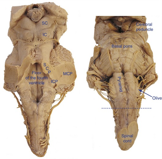
Various nuclei and fiber bundles form surface features at different levels of the brainstem. The most prominent of these are listed in this section (Fig. 11-1).
The Internal Structure of the Brainstem Reflects Surface Features and the Position of Long Tracts
The caudal or closed medulla is the part that does not contain any portion of the fourth ventricle (Fig. 11-2); it extends from the pyramidal decussation to the beginning of the fourth ventricle. The posterior columns start to terminate in nuclei gracilis and cuneatus in the caudal medulla; axons of these second-order neurons arch through the reticular formation as internal arcuate fibers, cross the midline, and turn upstream as the medial lemniscus. The rostral or open medulla is the part that contains a portion of the fourth ventricle (Fig. 11-3); it extends from the caudal end of the fourth ventricle to the point at which the brainstem becomes attached to the cerebellum by the inferior and middle cerebellar peduncles. The pyramids are still there, and now the inferior olivary nucleus gets added. Axons of these neurons curve across the midline as more internal arcuate fibers and form most (but not nearly all) of the inferior cerebellar peduncle, which turns up into the cerebellum right at the pontomedullary junction.
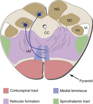
Figure 11-2 Caudal medulla (pyramids, central canal). As explained in Chapter 12, the spinal trigeminal tract and nucleus are the parts of the trigeminal system that take care of pain and temperature information from the head. CC, Central canal; IAF, internal arcuate fibers; NC, nucleus cuneatus; NG, nucleus gracilis; Vn, spinal trigeminal nucleus; Vt, spinal trigeminal tract.
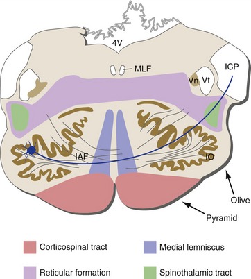
Figure 11-3 Rostral medulla (pyramids, fourth ventricle). 4V, Fourth ventricle; IAF, internal arcuate fibers; ICP, inferior cerebellar peduncle; IO, inferior olivary nucleus; MLF, medial longitudinal fasciculus (explained in Chapter 12); Vn, spinal trigeminal nucleus; Vt, spinal trigeminal tract.
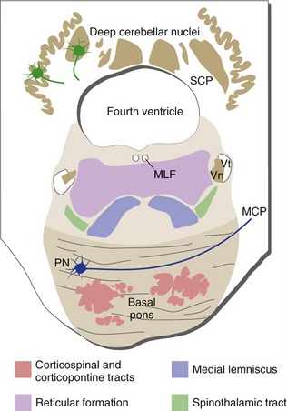
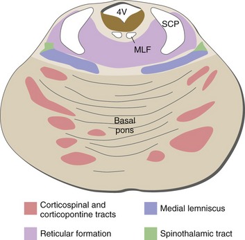
Every level of the pons contains part of the basal pons and fourth ventricle; the caudal pons is the part physically attached to the cerebellum, primarily by the middle cerebellar peduncles (Fig. 11-4). Here the medial lemniscus starts to flatten out and move laterally, and axons emerging from deep cerebellar nuclei start to form the superior cerebellar peduncle. The rostral pons is no longer connected to the cerebellum (Fig. 11-5); the middle cerebellar peduncles haven’t formed yet, and the superior cerebellar peduncles have left the cerebellum and are traveling rostrally through the brainstem. The trigeminal nerve is attached to the brainstem at the caudal pons-rostral pons junction.
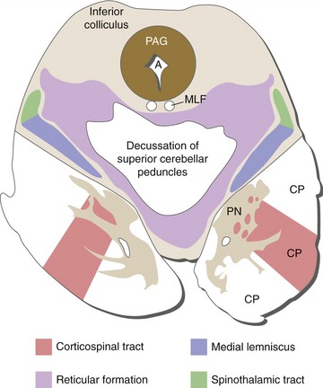
The cerebral aqueduct continues the ventricular system through the midbrain, surrounded by a distinctive area of periaqueductal gray that participates in many of the autonomic control functions discussed in Chapter 23. The caudal midbrain is the part containing the inferior colliculi (Fig. 11-6). Here the superior cerebellar peduncles decussate, so inputs to the cerebellum from each cerebral hemisphere cross in the basal pons and outputs cross back in this decussation of the superior cerebellar peduncles. The rostral midbrain is the part containing the superior colliculi (Fig. 11-7); it also contains two other distinctive areas of gray matter, the red nucleus and substantia nigra. The red nucleus is hooked up in cerebellar circuitry (see Chapter 20), and the substantia nigra is part of the basal ganglia (see Chapter 19). The trochlear nerve emerges at the pons-midbrain junction, and the posterior commissure is located at the midbrain-diencephalon junction.
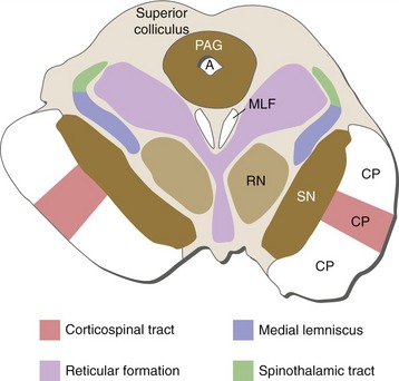
Figures 11-2 through 11-7 indicate just the major features of each of these brainstem levels. Information about additional brainstem functions and connections (e.g., cranial nerve nuclei) can be found in THB6 and in Chapters 12–14 of this book. The same figures are shown again in Chapter 15, with the structures discussed in Chapters 12–14 added in.
The Reticular Core of the Brainstem Is Involved in Multiple Functions
Some Brainstem Nuclei Have Distinctive Neurochemical Signatures
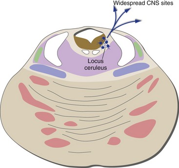
Neurons of the Locus Ceruleus Contain Norepinephrine
Neurons that use norepinephrine as a transmitter (noradrenergic neurons) are found in the peripheral nervous system as postganglionic sympathetic neurons. In the CNS, some are located in the medullary reticular formation, but most are pigmented neurons of the locus ceruleus in the rostral pons (Fig. 11-8). CNS noradrenergic neurons collectively project to practically every part of the CNS.
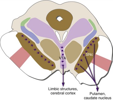
Neurons of the Substantia Nigra and Ventral Tegmental Area Contain Dopamine
Most of the neurons that use dopamine as a transmitter (dopaminergic neurons) are located in the midbrain (Fig. 11-9), either in the compact part of the substantia nigra or closer to the midline in the ventral tegmental area. Nigral dopaminergic neurons project to the caudate nucleus and putamen, and their degeneration causes Parkinson’s disease. Ventral tegmental neurons project to an assortment of limbic structures and (mostly frontal) cortical areas, and malfunction of these neurons or their targets plays a role in some forms of mental illness.
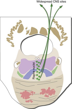
Neurons of the Raphe Nuclei Contain Serotonin
Neurons that use serotonin as a transmitter (serotonergic neurons) are located primarily in the raphe nuclei, a collective name for a series of nuclei near the midline of the brainstem reticular formation (Fig. 11-10). Like the noradrenergic neurons of the locus ceruleus, the serotonergic neurons of the raphe nuclei project practically everywhere in the CNS, suggesting that they too may be involved in adjusting levels of attention or arousal.
Neurons of the Rostral Brainstem and Basal Forebrain Contain Acetylcholine
Neurons that use acetylcholine as a transmitter (cholinergic neurons) play an important role in the peripheral nervous system. Some are located entirely in the periphery (postganglionic parasympathetic neurons), whereas others have their cell bodies in the CNS and axons that travel through spinal or cranial nerves (lower motor neurons, preganglionic sympathetic and parasympathetic neurons).
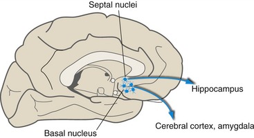
Some CNS cholinergic neurons are local interneurons in various structures (such as the putamen and caudate nucleus). Others have longer axons that project from one part of the CNS to another. Some of these are located in the midbrain reticular formation, but the largest collection is in the basal nucleus (of Meynert), a group of large cholinergic neurons in the basal forebrain (Fig. 11-11) that project to widespread areas of the cerebral cortex. Nearby cholinergic neurons in the septal nuclei project to the hippocampus. These basal forebrain neurons degenerate in victims of Alzheimer’s disease.
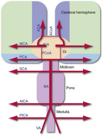
The Brainstem Is Supplied by the Vertebral-Basilar System
The vertebral arteries run along the lateral and anterior surfaces of the medulla and fuse to form the basilar artery, which runs along the anterior surface of the pons until it bifurcates into the posterior cerebral arteries on the anterior surface of the midbrain (THB6 Figures 6-3 and 11-29, pp. 126 and 291). Hence, nearly the entire supply of the brainstem comes from the vertebral-basilar-posterior cerebral system (THB6 Figure 11-30, p. 292). Knowing where each of the major branches from this system arises lets you surmise the supply of different levels of the brainstem (Fig. 11-12), because even if a branch (e.g., PICA) is headed for someplace like the cerebellum it needs to wrap around the brainstem to get there.