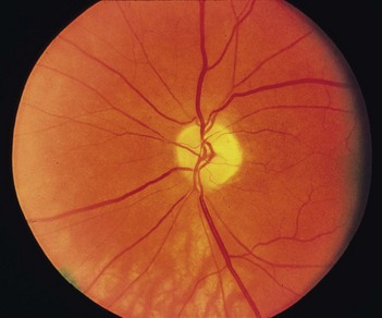
204 Optic atrophy
Salient features
Questions
What is the differential diagnosis?
• Demyelinating disorders (multiple sclerosis)
• Optic nerve compression by tumour or aneurysm
• Toxins: methanol, tobacco, lead, arsenical poisoning
• Ischaemia, including central retinal artery occlusion in thromboembolism, temporal arteritis, idiopathic acute ischaemic optic neuropathy, syphilis
• Hereditary disorders: Friedreich’s ataxia, Leber’s optic atrophy (sex linked, seen in young males)
Advanced-level questions
What is the difference between primary and secondary optic atrophy?
| Primary | Secondary |
|---|---|
| White and flat with clear-cut edges | Greyish-white, edges indistinct |
| Visible lamina cribrosa | Cup filled and lamina cribrosa not visible |
| Arteries and veins normal | Arteries thinner than normal, veins may be dilated |
| Capillaries decreased in number | Capillaries decreased in number (fewer than seven): Kestenbaum’s sign |
What is glaucomatous optic atrophy?
Glaucomatous optic atrophy denotes loss of disc substance, referred to as increased cupping.
How would you investigate a patient with optic neuropathy?
• Skull radiograph of pituitary fossa, optic foramina and sinuses, or CT scan of the brain and orbit


