Open Ventral Hernia Repair

Published on 16/04/2015 by admin

Filed under Surgery

Last modified 22/04/2025

Print this page

rate 1 star rate 2 star rate 3 star rate 4 star rate 5 star
Your rating: none, Average: 0 (0 votes)

This article have been viewed 1531 times

Chapter 31

Open Ventral Hernia Repair

Introduction

Ventral (abdominal) hernia repair is one of the most common procedures performed by general surgeons, with more than 200,000 done each year in the United States. Despite the prevalence of this procedure, there is little consensus as to the indications for repair, optimum technique, or appropriate position of the prosthetic mesh. Given the wide variety of patient and hernia factors, no single approach will likely suffice to repair all abdominal wall defects. Options include a laparoscopic or an open repair. The laparoscopic approach is typically reserved for obese patients with small to medium-sized defects, to avoid extensive subcutaneous dissection and potential mesh infections. The open repair can be performed with several methods.

Most surgeons agree that all incisional hernias should be repaired with prosthetic (synthetic or biologic) mesh, because recurrence rates are reduced by half. Prosthetic mesh can be placed as an inlay (sewn to the fascial edge), an onlay (sewn above the fascia), or a sublay (underneath the fascia). Sublay mesh can be placed in the intraperitoneal, preperitoneal, or retrorectus position. The inlay approach has been largely abandoned because of high recurrence rates, and the onlay approach is discouraged because the prosthetic mesh is placed in the subcutaneous position at highest risk for mesh sepsis. Most herniologists agree that the prosthetic mesh should be placed as a sublay. This chapter focuses on the sublay repair, with particular attention to the retrorectus placement of the mesh.

Surgical Principles

General principles of incisional hernia repair include gaining access to the reoperative abdomen, complete adhesiolysis, preparation of the abdominal wall for prosthetic mesh placement, mesh deployment, and reconstruction of a dynamic abdominal wall with reapproximation of the linea alba. Safe access to the reoperative abdomen typically involves extending incisions in a cephalad direction to enter the undissected peritoneum. Complete adhesiolysis of the anterior abdominal wall is important to free the abdominal wall musculature and allow it to advance to the midline during eventual reconstruction of the midline.

Reestablishing the linea alba is an important concept in abdominal wall reconstruction. If the linea alba is seen as the tendinous insertion of the rectus abdominis muscle and oblique muscles, it is critical to achieve appropriate physiologic loading of the abdominal wall. In particular, reconstructing a completely tension-free repair in fact renders the oblique muscles nonfunctional, with constant lateral displacement of the abdominal wall leading to mesh displacement at the mesh-tissue interface. This chapter emphasizes surgical methods to reapproximate the rectus complex.

Preoperative Imaging

Preoperative imaging of the abdominal wall is very helpful. The author typically performs abdominal pelvic computed tomography (CT) scans in all patients undergoing complex abdominal wall reconstruction. CT images can help delineate the location, size, and complexity of the hernia, particularly in obese patients. Attention to the relationship of the hernia to bone structures (e.g., xiphoid, costal margin, iliac crest, pelvis) is important for surgical planning. Recognizing the appropriate plane is paramount to gain sufficient overlap of the mesh and achieve the most durable repair. Likewise, the integrity of the rectus abdominis muscle is important. If the rectus muscle is narrow, the mesh cannot be placed in the retrorectus position and will need to be located in the preperitoneal position, as described later. If the rectus muscle is destroyed, absent, or atrophic, alternative methods are necessary.

Some surgeons have advocated abdominal wall ultrasound to detect hernias, but the author has found this to be very user dependent, with minimal experience.

Anatomy of Abdominal Wall

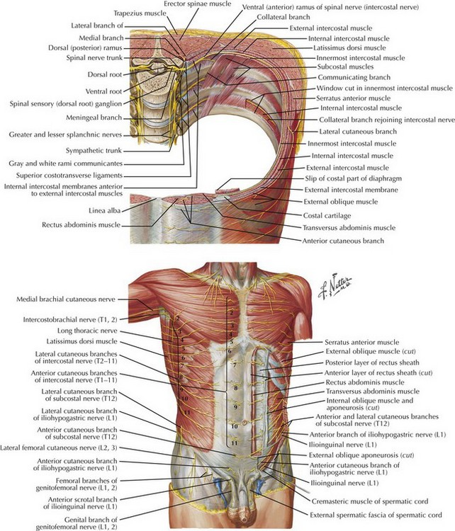
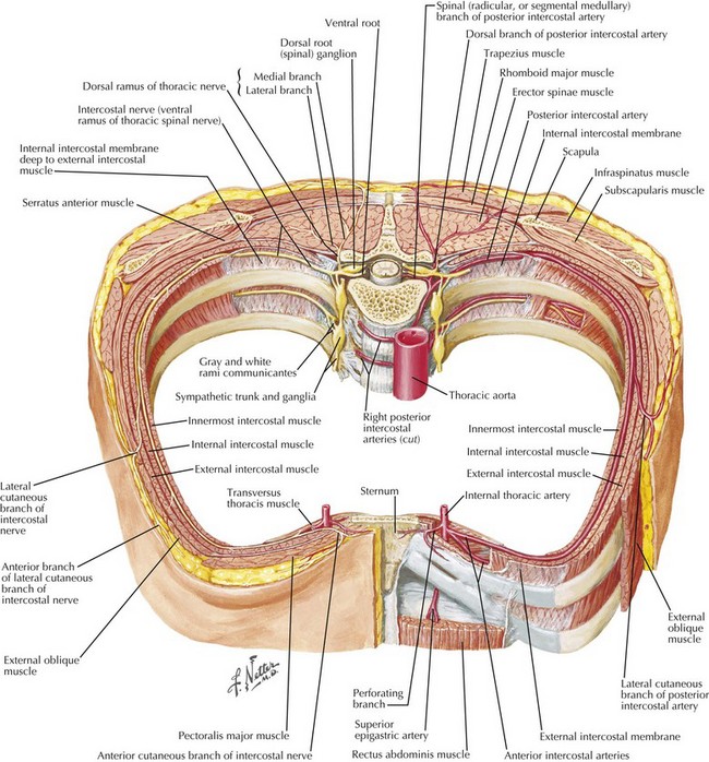
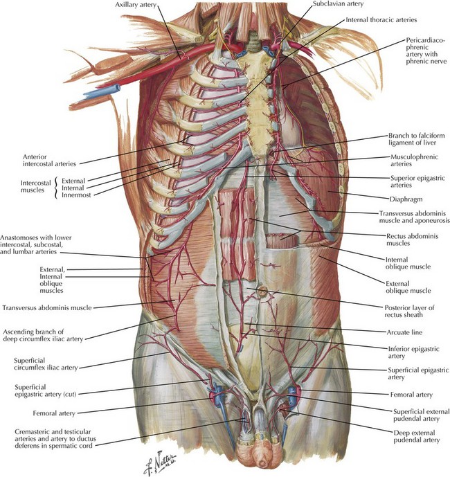
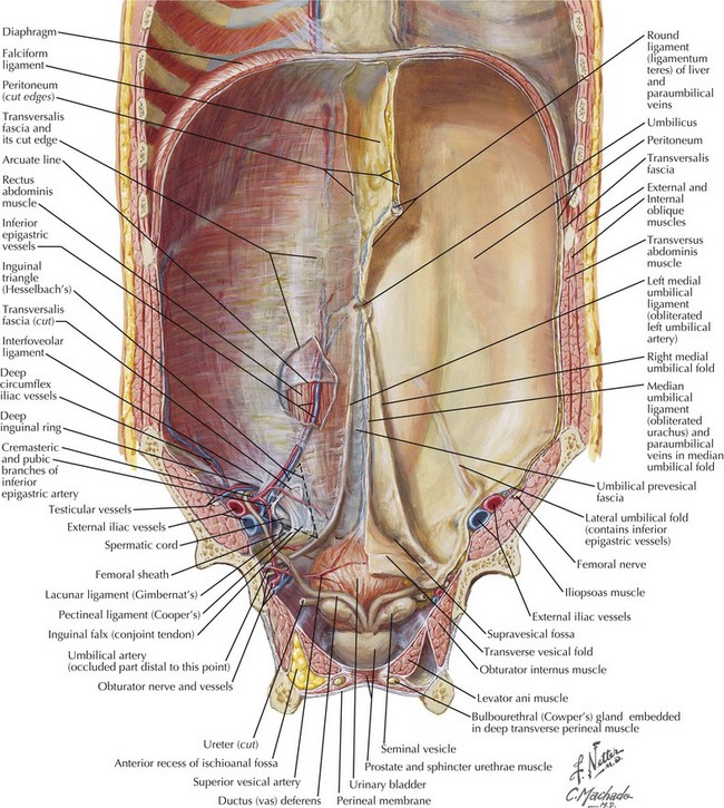
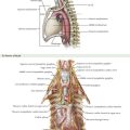
Figures 31-1 and 31-2 show the anatomy of the vascular supply and innervation of the abdominal wall. Understanding the relationships of these nerves and vessels and their location in the abdominal wall is critical to preserve them during dissection, to maintain an innervated functional abdominal wall.

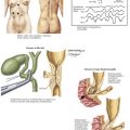
The blood supply of the anterior abdominal wall can be divided into three zones (Fig. 31-3). Zone 1 consists of the upper and midcentral abdominal wall and is supplied by the deep superior and deep inferior epigastric arteries. Zone 2 consists of the lower abdominal wall and is supplied by the epigastric arcade and the superficial inferior epigastric, superficial external pudendal, and superficial circumflex iliac arteries. Zone 3 consists of the lateral abdominal wall (flank) and is supplied by the musculophrenic and lower intercostal and lumbar arteries. Recognizing the location of prior transverse incisions that may have compromised abdominal wall blood supply is important to limit ischemic skin complications.

Sensory innervation of the abdominal wall is derived from the 7th thoracic (T7) to 1st lumbar (L1) intercostal and subcostal nerves. These nerves run alongside the intercostal and lumbar arteries in the plane between the internal oblique and transversus abdominis muscles. The rectus abdominis muscle is segmentally innervated by the lower six intercostal nerves. These nerves penetrate the linea semilunaris at the lateral border of the rectus muscle. It is important to preserve these nerves during dissection of the lateral abdominal wall, to avoid denervation of the rectus complex.

Surgical Technique

A standard midline incision is created to encompass all previous surgical incisions, if possible. The abdomen is entered sharply, and the anterior abdominal wall is completely freed of adhesions to the lateral gutters (adhesiolysis). It is important to separate all adhesions to avoid injuring visceral contents during dissection of the lateral abdominal planes, and to allow these structures to slide to the midline during eventual abdominal wall reconstruction. A surgical towel is placed over the viscera to protect them during dissection. All prior prosthetic materials are removed from the abdominal wall.

Creation of Retrorectus Space

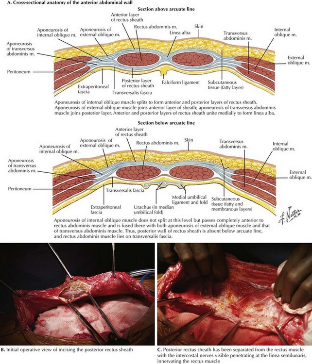
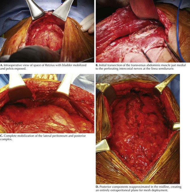
The linea alba is grasped with Kocher clamps, and the posterior rectus sheath is incised approximately 0.5 cm lateral from its edge (Fig. 31-4, A and B). This typically is begun just above the umbilicus. The plane is created using cautery, with care taken to avoid injuring the underlying rectus muscle. The retromuscular plane is then developed in a cephalad and caudal direction. The dissection is carried laterally to the linea semilunaris. This anatomic plane is localized by identifying the perforating intercostal nerves and vessels (Fig. 31-4, C). It is typically 1 cm lateral to the inferior epigastric vessels.

If the rectus muscle is relatively well preserved and sufficiently wide, the dissection is complete; the posterior components are closed and prosthetic mesh is placed. In larger hernias, requiring more overlap, or in atrophic narrowed rectus muscles, the dissection can be continued to the lateral abdominal wall (see Lateral Dissection in Preperitoneal Plane).

Exposure of Cooper’s Ligament and Pelvis

The dissection can be continued inferiorly to the pubis, if necessary (Figs. 31-5 and 31-6, A). The space of Retzius is entered bluntly to expose the pubic symphysis in the midline. If undissected, this plane is typically bloodless and can be easily developed. If the bladder has previously been mobilized, using a three-way Foley catheter with instillation of 300 mL of saline into the bladder can aid in identifying and avoiding a bladder injury. This plane is below the arcuate line and therefore consists of the peritoneum and transversalis fascia only. Fenestrations can occur and should be recognized and repaired accordingly. The pubis and bilateral Cooper’s (pectineal) ligament are exposed. The inferior epigastric vessels, iliac vessels, and cord structures should be identified and carefully preserved in this dissection plane.

Lateral Dissection in Preperitoneal Plane

In patients with insufficient mobilization of the posterior sheath to reapproximate in the midline, narrow atrophied rectus muscles, and inability to place wide prosthetic mesh, the dissection is carried into the lateral abdominal plane. Ideally, this plane is entered while the innervation of the rectus complex is preserved. The preperitoneal space is entered approximately 1 cm medial to the perforating nerves at the linea semilunaris (Fig. 31-6, B and C). The posterior rectus sheath is divided throughout its length. With the use of blunt Kitner dissection, the preperitoneal space is developed to the psoas muscle, if necessary. This preperitoneal plane is particularly useful for subxiphoid hernias because the peritoneum can be swept off the costal margin several centimeters superiorly. As the dissection continues cephalad, the posterior sheath typically consists of the transversus abdominis muscle. This muscle must be divided to enter the preperitoneal space.

Posterior Layer Reconstruction

Once the release is completed on both sides, the posterior components are reapproximated in the midline, completely excluding the bowel from the mesh (Fig. 31-6, D).This procedure is performed with a running absorbable suture. Defects in the posterior layer are closed with interrupted figure-of-eight sutures or buttressed with native tissue (omentum, fat). Antibiotic irrigation is typically performed.

Mesh Deployment

The prosthetic mesh is placed in the sublay retromuscular position. Because the mesh is placed extraperitoneally, extensive sutures are not necessary because the bowel cannot herniate around the mesh. Likewise, a standard macroporous mesh is sufficient without an antiadhesive barrier. Full-thickness transfascial sutures are placed. These sutures serve two key purposes: providing stability to the mesh immediately after the repair to prevent displacement and helping medialize the rectus muscles and redistribute the tension of the repair to the lateral abdominal wall through the mesh. It is important to understand this concept, because this is not meant to be a true tension-free hernia repair. Instead, it restores appropriate physiologic tension to the abdominal wall. If the mesh is placed without tension, and the abdominal wall is then reapproximated, there is almost always some buckling of the mesh, which leads to seromas, infections, and lack of incorporation.

The inferior edge of the mesh can be secured directly to Cooper’s ligament. If the defect is suprapubic, the sutures are backed off the edge of the mesh, to allow the prosthetic material to drape below the pelvis. Suction drains are placed in the retromuscular space, above the mesh. These drains are removed rapidly if synthetic mesh is chosen, or are left for several weeks if biologic mesh is used.