[level-membership-for-opthalmology-category]12.4
Oculocutaneous Albinism
Clinical Features:
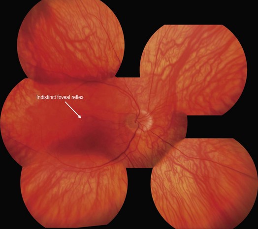
The fundus of tyrosinase-negative individuals have a complete lack of pigmentation, whereas tyrosinase-positive individuals have a variable, but reduced, amount of fundus pigmentation (Fig. 12.4.1). Foveal hypoplasia is characteristically present in both types.
OCT Features:
OCT line scans of the central macula reveal lack of a distinguished foveal depression, evidence of foveal hypoplasia (Fig. 12.4.2). As a result of this, the corresponding thickness map shows central ‘thickening’ in comparison to the normative database.
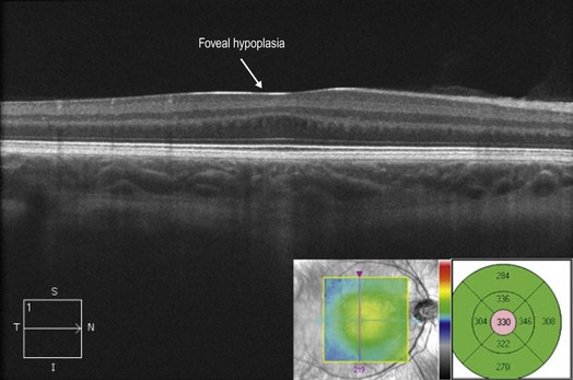
Figure 12.4.2 OCT line scan (corresponding to Figure 12.4.1) through the central macula shows foveal hypoplasia with lack of a well-defined foveal depression. The accompanying thickness map (inset) shows increased thickness centrally in comparison to a normative database due to the lack of a normal foveal depression.
[/level-membership-for-opthalmology-category][not-level-membership-for-opthalmology-category]12.4
Oculocutaneous Albinism
Clinical Features:
The fundus of tyrosinase-negative individuals have a complete lack of pigmentation, whereas tyrosinase-positive individuals have a variable, but reduced, amount of fundus pigmentation (Fig. 12.4.1). Foveal hypoplasia is characteristically present in both types.
[/not-level-membership-for-opthalmology-category]
