4 Obstetric examination
Abdominal examination
Palpation
• Is the fetus appropriately grown for gestation? The symphyseal–fundal height of the uterus (in centimetres) should correspond with the gestation + 1–2 weeks.
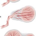
• Does the liquor volume feel normal? This could be considered in a larger-for-dates tense uterus in which the fetus is difficult to palpate (polyhydramnios) or, conversely, in a smaller-for-dates uterus in which very little fluid is palpated around the fetus (oligohydramnios).
• Is this a singleton or multiple pregnancy, i.e. greater than two fetal poles can be palpated?
• Is the lie of the fetus (the relation of the fetal spine to the longitudinal axis of the uterus) longitudinal or oblique or transverse?
• Is the presentation cephalic or breech? If cephalic, is it engaged or how palpable is it above the pelvis?
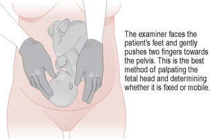
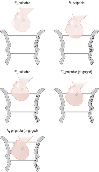
To measure the symphyseal–fundal height, feel for the fundus of the uterus with the tips of the fingers or ulnar surface of the hand and measure the distance to the symphysis pubis with a tape measure, turning it over so that the centimetre markings cannot be seen, to avoid bias (Fig. 4.1). The manoeuvres for palpation of the uterus include: lateral uterine grip with the hands, assessing the lie of the fetus by palpating the sides of the uterus (Fig. 4.2); fundal grip with the hands, assessing the fetus at the uterine fundus; the deep pelvic grip with both hands pointed towards the pelvis to assess for presentation and engagement, ensuring that one is not causing discomfort by observing the mother’s face during palpation. An alternative to the latter is Pawlik’s grip, which is the grasping of the presenting fetal part between the index finger and thumb, taking care to avoid patient discomfort. Engagement of the fetal head is said to occur when the largest diameter has passed into the pelvis. For a subjective description of this, the fetal head is divided into fifths palpable abdominally. The head is engaged if less than two-fifths of it can be palpated abdominally (Fig. 4.3).
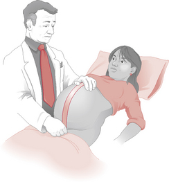
Figure 4.1 Measuring the height of the fundus.
(Redrawn with permission from Hanretty K P 2003 Obstetrics Illustrated, 6th edn. Edinburgh: Churchill Livingstone, p. 76.)