Nutritional Dermatoses

Published on 06/06/2015 by admin

Filed under Pediatrics

Last modified 22/04/2025

Print this page

rate 1 star rate 2 star rate 3 star rate 4 star rate 5 star
Your rating: none, Average: 0 (0 votes)

This article have been viewed 3503 times

127 Nutritional Dermatoses

Appropriate nutritional intake is an important aspect in children’s overall health. Although health care workers in developing countries are confronted daily with patients with various forms of nutritional deficiencies, these important health issues can also manifest in affluent countries with abundant food supplies. Infants and children require sufficient calories and nutrients for normal growth and development. Malnutrition in general is considered by the World Health Organization to be one of the most important risk factors for illness and premature death.

Clinical Presentation
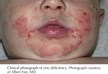
Protein-Energy Malnutrition

The majority of nutritional deficiency is associated with either poor dietary intake or GI malabsorption. Protein-energy malnutrition is divided into two major categories, marasmus and kwashiorkor. Marasmus is caused by insufficient total caloric intake and classically is seen in the context of food deprivation. Conversely, kwashiorkor is associated with inadequate protein intake in the setting of normal caloric intake. Indeed, kwashiorkor was initially described in children whose caloric intake was almost entirely derived from corn. Protein-losing enteropathies or diets consisting entirely of rice, milk because of concerns for milk intolerance are known causes of kwashiorkor in developed countries.

A child with marasmus is defined as having less than 60% of expected body weight for age in the absence of edema or hypoproteinemia. This form of malnutrition presents with loss of subcutaneous fat and muscle wasting, leading to an overall emaciated appearance. The skin is typically dry, thin, and wrinkled. Hyperpigmentation, fine scale, increased lanugo hair, nail fissures, and purpura are other cutaneous findings associated with marasmus.

Children with kwashiorkor also exhibit a lower body weight than expected for age, ranging from 60% to 80% of expected body weight. However, unlike in marasmus, these children also exhibit edema and hypoproteinemia stemming from insufficient dietary protein or the underlying medical condition leading to intestinal protein loss. Overall, the presence of mild hypoproteinemic edema in early disease gives the appearance of a well-fed, overweight child. However, recognition of the cutaneous findings help establish the diagnosis. Pigment alterations are the most common skin finding in patients with kwashiorkor, presenting as hypo- or hyperpigmentation after minor injury. In mild cases, superficial desquamation occurs that has the clinical appearance of enamel paint, which can progress in more severe cases to large areas of erosions, particularly on the extremities and buttocks, with an appearance similar to flaking paint. These areas are commonly secondarily infected with bacteria or colonized with Candida spp. Children also can present with skin atrophy, redness, and purpura. The hair is often dry, sparse, and lighter in color. Under conditions of repeated episodes of protein malnutrition followed by periods of adequate protein intake, the hair may have alternating light and dark bands, termed the flag sign, which correspond to the different episodes of protein nutritional status. Other important findings helpful in establishing the diagnosis of kwashiorkor include edema, irritability, anorexia, apathy, hepatomegaly from fatty infiltration of the liver, and failure to thrive.

Zinc Deficiency

Zinc is an element that is a required component of many enzymes involved in the synthesis and degradation of lipids, protein, and nucleic acid. Classic skin findings of zinc deficiency include erythematous and slightly eroded plaques involving the extremities, diaper area, and periorificial area. The facial involvement often involves the lower cheeks and chin but spares the skin above the upper lip, giving a “U” appearance. The rash can present with exudate, crust, vesicles, and bullae (Figure 127-1). Chronic zinc deficiency often manifests with lichenified plaques. Candida spp. and Staphylococcus aureus superinfections are common in this disorder. Other cutaneous findings of zinc deficiency include stomatitis, angular chelitis, blepharitis, nail-fold inflammation with possible nail dystrophy, and hair thinning with areas of complete alopecia. In addition to dermatitis, diarrhea is a commonly associated symptom. The severity is highly variable and does not correlate with the development of cutaneous findings. Children with zinc deficiency also characteristically are irritable, have problems eating and sleeping, and are growth impaired.
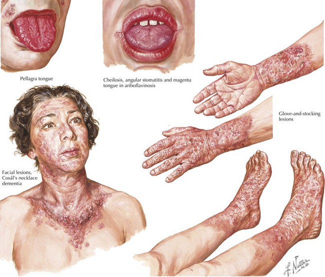
Acquired zinc deficiency is associated with poor dietary intake or underlying GI disease, leading to malabsorption. In addition to acquired zinc deficiency, an inherited cause of zinc deficiency has been identified. This disorder, termed acrodermatitis enteropathica, is caused by an autosomal recessive mutation in the zinc transporter gene SLC39A4. Because human breast milk (but not formula or cow’s milk) contains zinc-binding proteins that aid in zinc absorption, these patients typically present 1 to 2 weeks after weaning from breast milk. Interestingly, various case reports of exclusively breastfed infants with zinc deficiency demonstrated decreased zinc levels in breast milk despite normal maternal serum zinc. These infants do not exhibit a defect with intestinal zinc absorption, classifying this disorder, termed transient neonatal zinc deficiency, as an acquired zinc deficiency unlike acrodermatitis enteropathica. Transient neonatal zinc deficiency has been linked with maternal mutations in the zinc transporter gene SLC30A2, which is responsible for transport of zinc into breast milk. Understanding the genetic cause of zinc deficiency is important because the levels of zinc supplementation for acquired zinc deficiency and acrodermatitis enteropathica differ, and zinc oversupplementation can potentially result in immune dysfunction. The diagnosis of other specific nutritional deficiencies, including essential fatty acid (EFA) deficiency and biotin deficiency, should be considered when suspecting zinc deficiency because of the similarity in cutaneous manifestations of these three disorders (see below).

Biotin Deficiency

Biotin, which is also known as vitamin H or vitamin B7, is a water-soluble vitamin B complex nutrient that functions as a cofactor for carboxylases, which are enzymes involved in gluconeogenesis and the metabolism of amino acids and fatty acids. Biotin deficiency occurs in children receiving total parenteral nutrition without biotin supplementation. Unusual diets consisting of many raw eggs can lead to biotin deficiency because of high levels of the protein avidin in raw egg whites that binds and inactivates biotin. Biotin is also produced by bacteria in the large intestines, so broad-spectrum antibiotics may trigger a biotin deficiency because of altered intestinal flora. Other drugs, particularly anticonvulsants such as phenytoin, phenobarbital, and carbamazepine, have the potential to cause biotin deficiency because of their impairment of biotin absorption.

The inherited form of biotin deficiency is called multiple carboxylase deficiency. Two distinct genes have been associated with this disorder, holocarboxylase synthetase and biotinidase, two enzymes required for utilization of biotin. Holocarboxylase synthetase deficiency typically presents during infancy, and biotinidase deficiency presents in children between 3 months of age and 2 years of age. Recently, a case report of a child with multiple carboxylase deficiency despite normal dietary biotin intake and normal holocarboxylase synthetase and biotinidase suggests that a biotin transport protein deficiency can also cause biotin deficiency.

Cutaneous findings of biotin deficiency include a periorificial dermatitis resembling zinc deficiency, alopecia, blepharitis, and conjunctivitis. Organic aciduria; developmental delay; and neurologic findings such as seizures, ataxia, and hypotonia are also seen in children with biotin deficiency. Children also are at increased risk of fungal infections because of decreased immune function. In neonates, holocarboxylase synthetase deficiency often initially presents with metabolic acidosis, and without prompt recognition of the diagnosis and appropriate supplementation, it can lead to death. Although the cutaneous findings associated with all types of biotin deficiency can be reversed with supplementation, the neurologic impairment related to biotin deficiency may be permanent, further supporting the need for recognition of this deficiency.

Niacin Deficiency

Niacin is a water-soluble nutrient that is part of the vitamin B complex. Deficiency of niacin, also known as vitamin B3, leads to pellagra. The classic skin manifestations of pellagra are symmetric erythema typically occurring on sun-exposed sites associated with itching and burning that can form blisters and crusts after sunlight exposure. The lesions later become coarse, dry, and hyperpigmented. On the upper chest, keratotic hyperpigmented plaques associated with pellagra are commonly known as Casal’s necklace. Oral findings include edema, atrophic glossitis, and stomatitis (Figure 127-2). Patients with niacin deficiency also develop GI symptoms, such as abdominal pain, vomiting, and diarrhea, and neurologic complaints, such as vertigo. Although the symptoms of pellagra are often summarized into the three Ds—dermatitis, diarrhea, and dementia—the full spectrum of the disease is not usually seen during the early stages and is rare in pediatric patients. Although niacin is found in most foods, it is not absorbed well from corn, and thus children whose diet is almost entirely corn are at high risk of developing pellagra. Tryptophan can be converted to niacin in the body; therefore, conditions associated with tryptophan malabsorption increase the risk of developing pellagra-like symptoms. Hartnup’s disease is a genetic condition in which patients have a genetic mutation in the neutral amino acid transporter gene (SLC6A19), resulting in poor intestinal tryptophan absorption and the potential for developing symptoms of pellagra. Drugs such as isoniazid and 5-fluorouracil that inhibit the conversion of tryptophan to vitamin B3 or phenytoin that can decrease total serum tryptophan also increase susceptibility to development of pellagra.
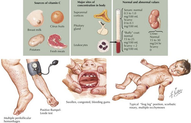
Other Vitamin B Complex Deficiencies

Vitamin B6 is also known as pyridoxine, which is converted in the body into pyridoxal-5-phosphate, a coenzyme important in amino acid metabolism. Deficiency of vitamin B6 manifests with oral changes typical of all vitamin B complex deficiency, including glossitis and stomatitis. A dermatitis similar to pellagra can also be observed, likely because vitamin B6 is a cofactor in the enzyme important for the conversion of tryptophan to niacin. Vitamin B6 deficiency can manifest as a periorificial, scaly rash that resembles seborrheic dermatitis.

Deficiency of vitamin B12 (cyanocobalamin) and folic acid (vitamin B9) have similar clinical findings, and the most common is megaloblastic anemia. Cutaneous findings include painful atrophic glossitis and angular chelitis similar to many other vitamin B complex deficiencies. Dark-skinned patients may also present with hyperpigmentation with a predilection for the flexural areas, creases of the palms and soles, and nails. Many foods are fortified with folic acid, making isolated deficiency rare. Vitamin B12 is important in DNA synthesis, so children with various chronic anemias such as sickle cell disease have increased folic acid requirements. Vitamin B12 deficiency is rare in infants because the maternally derived stores last approximately 1 year. A strict vegetarian diet (veganism) is associated with vitamin B12 deficiency as well as decreased intestinal absorption caused by gastric intrinsic factor deficiency (pernicious anemia).