Nummular eczema
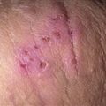
Nummular eczema may look like ringworm or psoriasis. The borders of ringworm and psoriasis are usually sharply defined. The lesion borders are indistinct in this case of nummular eczema.
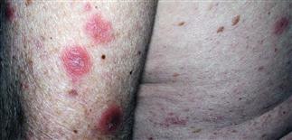
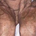
The most common areas are the dorsa of the hands, the lower legs, the upper extremities (shown here), and the trunk. Men are most commonly affected.
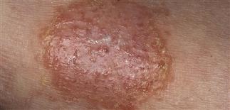
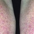
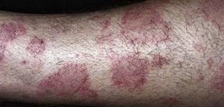
Sharply circumscribed erythematous plaque with weeping surface. Nummular dermatitis may be a solitary lesion, but more frequently there are multiple similar lesions.
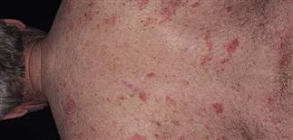
On the back there are scattered erythematous plaques which are intensely pruritic, leading to poor quality of life. This nummular eczema may last many months and may be refractory to treatment.
DESCRIPTION
Round (coin-shaped) lesions of eczematous inflammation, often chronic, treatment-refractory. Cause unknown, occasionally allergic contact allergens are relevant. Intensely itchy.
HISTORY
• Adults most often affected, men more than women. • Onset usually gradual, with no clear precipitant and no eczema history. • Often begins with few isolated lesions on legs; with time, multiple lesions occur, without particular distribution. • Lesions often resolve or improve after use of topical corticosteroids but recur in same area after steroid withdrawal. Can become refractory even to high-strength topical steroids. • Course variable, unpredictable; may be chronic and relapsing for years. • Once lesions established, they tend to remain same size and recur on previously affected skin. • Differential diagnosis includes psoriasis, tinea, cutaneous T-cell lymphoma.
PHYSICAL FINDINGS
• Sharply demarcated, round eczematous plaques on trunk and extremities. • Weeping lesions and vesiculation characterize flares. • Secondary infection can result in disease flares. • A yellow, honey-colored crust indicates secondary impetiginization. • Patch testing reveals a relevant positive result in small percentage of cases (one-quarter to one-third). • Culture may reveal Staphylococcus aureus. Antibacterial treatment usually helps but does not often resolve problem.
TREATMENT
• See Subacute eczematous inflammation or Chronic eczematous inflammation. • Consider patch testing in refractory cases. • Discontinue unnecessary routine topical moisturizing products, over-the-counter oral medications, dietary supplements, herbal preparations for at least 3–4 months. • Active tinea pedis should be sought; treat tinea with antifungal agents if present. • Use of medium-potency topical steroid ointments and bland emollients (Aquaphor, Vanicream, Vaseline) should be aggressive. • Apply topical steroid twice a day for 2–3 weeks. Treat a week or so longer than deemed necessary to resolve lesion. • Efficacy of topical steroid increased by occlusion with plastic wrap or sauna suit, or by hydrating skin with a bath before applying medication. • Treat secondary infection with systemic antistaphylococcal antibiotics, according to sensitivity results (e.g. dicloxacillin 250 mg q.i.d., cephalexin 250 mg q.i.d.). • Antihistamines can be prescribed for itching. • Systemic corticosteroids should be avoided for long-term management. • Refer refractory cases to a dermatologist. • Light therapy can help resolve lesions when topical treatments have failed. Narrow- and broadband ultraviolet B are best choices for light therapy; can use psoralen plus ultraviolet A if ultraviolet B therapy fails. • Topical allergens such as fragrance and medicaments (bacitracin, Neosporin, hydrocortisone) may drive some cases of nummular dermatitis. Counsel against their use.