[level-membership-for-radiology-category]
16 Nuclear medicine
Definition of nuclear medicine
| The introduction of a specific pharmaceutical (depending on which part of the body is to be targeted), which has been labelled with a radioisotope, into a patient. The gamma rays emitted by the radioisotope are scanned by a detector and the diagnostic image is produced showing the concentrations of the radiopharmaceutical (e.g. a bone scan) or an indication of function (e.g. the glomerular filtration rate of the kidneys) |
| Charge Collection | The pooling of electrons across a crystal |
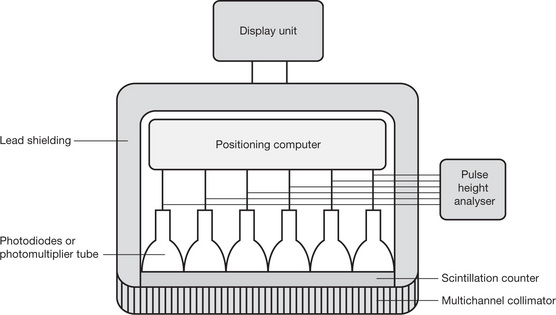
| Gamma Camera | A large, stationary, scintillation counter, which records the activity over the whole field at the same time. Used to detect pathologies where the physiology of the structure is changed |
| Gantry | A structure or support, in which the X-ray tube, detectors and associated electronics are housed |
| Half-life | The amount of time taken for the radioactivity of a radioactive substance to decay by half the initial value. The half-life is a constant for each radioactive isotope |
| Image Fusion | When a PET (or SPECT) image and a CT image are viewed together by one being superimposed on the other |
| Pharmaceutical | A drug used in medicine |
| Photodiode | A semiconductor used to detect light and then generate electricity in proportion to the quantity of light detected |
| Photomultiplier | Equipment that produces an amplified current when exposed to electromagnetic radiation (light). Photons hitting the cathode produce electrons which in turn hit other surfaces thus producing more electrons, forming a pulse of electricity which forms the subsequent image |
| Planar | A two dimensional image |
| Pulse Height Analyser | Receives the signal from the photomultiplier and only produces an electrical signal if the input pulse lies in a predetermined range |
| Radioisotope | Any isotope that is radioactive. Forms of an element which have the same atomic number but different mass numbers, exhibiting the property of spontaneous nuclear disintegration |
| Radiopharmaceutical | A drug consisting of a radioactive compound |
| Scintillation Counter | A number of scintillator crystals in containers, one surface of the crystal is attached to a transparent glass window and the other surfaces are coated with magnesium oxide to reflect light back into the crystal; the back of the crystal is attached to a photomultiplier tube. If a gamma ray hits the crystal, light is produced and some reaches the photocathode of the photomultiplier |
| Scintillator | A sodium iodide (or caesium iodide) crystal with a thallium activator |
| Segmented | Divided into sections |
| Solid State Detector Material | Semi conductor – Cadmium zinc telluride (CdZnTe) |
| Spatial Resolution | The smallest distance between two objects that can be visually seen on an imaging system |
| Pharmaceutical | A drug which is absorbed by a specific (targeted) area of the body |
| Radioisotope | Technetium 99m (99Tcm) most commonly used
It is combined with a specific pharmaceutical so that a specific area of the body will take up the radioisotope |
| Detector – Gamma Camera | Radiation from the patient passes through: A multichannel collimator A scintillation counter Photodiodes or photomultipliers Pulse height analyser |
| Operator Console | Where the operator can determine the settings for the scan |
| Display Station | For the viewing, analysis, networking and storage of the final image |
| Rectilinear Scanners | |
| Gamma Cameras | |
| Mobile Gamma Cameras | |
| Dual Headed Gamma Cameras (GCPET) | |
| Advantages | |
| Disadvantages | |
| Gamma Probe | Designed for use by a surgeon to detect small quantities of tissue labelled with either Technetium 99m or Indium 111
• The tube is shielded with tungsten and has clip-on collimators to reduce the radiation to the operator
Application |
Positron Emission Tomography (PET)
| Definition | When a positron is emitted it travels a few millimetres then annihilates with a free electron resulting in the emission of two 511 keV photons leaving at nearly 180° to each other. A ring of scintillation detectors are positioned so that they capture the photons and produce a computerised image. Only if two detectors opposite each other register a photon within a nanosecond of each other are the photons registered. They are used in conjunction with CT scanners where the CT scanner shows the anatomy and the PET scanner the function of an organ or tumour, the images being superimposed on each other. Planar or three dimensional images can be produced |
| Cyclotron | |
| Function | |
| Detectors | |
| Gantry | |
| Software | |
| Radioisotope | Examples |
| Radiopharmaceutical | Fluorine-18 fluorodeoxyglucose (18F-FDG) |
| Application |


Single Photon Emission Computerised Tomography (SPECT)
| Definition | A specialist gamma camera which rotates round the patient at 2.8 revolutions per minute and a number of two dimensional images with a slice thickness of 10 mm are taken by measuring the emission of single photons. They can be used in conjunction with CT scanners where the CT scanner shows the anatomy and the SPECT scanner the function of an organ or tumour, the images being superimposed on each other |
| Gamma Camera | |
| Gantry | |
| Software | |
| Radioisotope | Examples |
| Radiopharmaceutical | Examples |
| Application | |
| Limitations |


| Dakin M 2001 Positron emission tomography in the UK. Synergy, November | |
| Griffiths M 2005 SPECT/CT hybrid imaging technology, techniques and clinical experience. Synergy, January | |
| Griffiths M, Aston A, Roberts F 2003 CT 2003 future’s bright the future’s fusion. Synergy, November | |
| Griffiths M, Holmes K 2002 The development of nuclear medicine equipment. Synergy, November | |
| Higgins R, Smith L, Vinjamuri S 2004 Pancreatic carcinoma and imaging with PET. Synergy, May | |
| Hogg P, Lewington G 2005 An overview of PET/CT and its place in today’s UK healthcare system. Synergy, December | |
| Millns M, Owens S, 2001 Unclear nuclear medicine? Not any more! Synergy, February | |
| Moorhouse S 2005 Gamma camera SPET imaging of solitary pulmonary nodes. Synergy, January | |
| Old S E, Dendy P P, Balan K K 2000 Preliminary experience in oncology of positron emission tomography with dual headed gamma camera. Radiography 6:11–17 |
[/level-membership-for-radiology-category][not-level-membership-for-radiology-category]
16 Nuclear medicine
Definition of nuclear medicine
| The introduction of a specific pharmaceutical (depending on which part of the body is to be targeted), which has been labelled with a radioisotope, into a patient. The gamma rays emitted by the radioisotope are scanned by a detector and the diagnostic image is produced showing the concentrations of the radiopharmaceutical (e.g. a bone scan) or an indication of function (e.g. the glomerular filtration rate of the kidneys) |
| Charge Collection | The pooling of electrons across a crystal |
| Gamma Camera | A large, stationary, scintillation counter, which records the activity over the whole field at the same time. Used to detect pathologies where the physiology of the structure is changed |
| Gantry | A structure or support, in which the X-ray tube, detectors and associated electronics are housed |
| Half-life | The amount of time taken for the radioactivity of a radioactive substance to decay by half the initial value. The half-life is a constant for each radioactive isotope |
| Image Fusion | When a PET (or SPECT) image and a CT image are viewed together by one being superimposed on the other |
| Pharmaceutical | A drug used in medicine |
| Photodiode | A semiconductor used to detect light and then generate electricity in proportion to the quantity of light detected |
| Photomultiplier | Equipment that produces an amplified current when exposed to electromagnetic radiation (light). Photons hitting the cathode produce electrons which in turn hit other surfaces thus producing more electrons, forming a pulse of electricity which forms the subsequent image |
| Planar | A two dimensional image |
| Pulse Height Analyser | Receives the signal from the photomultiplier and only produces an electrical signal if the input pulse lies in a predetermined range |
| Radioisotope | Any isotope that is radioactive. Forms of an element which have the same atomic number but different mass numbers, exhibiting the property of spontaneous nuclear disintegration |
| Radiopharmaceutical | A drug consisting of a radioactive compound |
| Scintillation Counter | A number of scintillator crystals in containers, one surface of the crystal is attached to a transparent glass window and the other surfaces are coated with magnesium oxide to reflect light back into the crystal; the back of the crystal is attached to a photomultiplier tube. If a gamma ray hits the crystal, light is produced and some reaches the photocathode of the photomultiplier |
| Scintillator | A sodium iodide (or caesium iodide) crystal with a thallium activator |
| Segmented | Divided into sections |
| Solid State Detector Material | Semi conductor – Cadmium zinc telluride (CdZnTe) |
| Spatial Resolution | The smallest distance between two objects that can be visually seen on an imaging system |
| Pharmaceutical | A drug which is absorbed by a specific (targeted) area of the body |
| Radioisotope | Technetium 99m (99Tcm) most commonly used
It is combined with a specific pharmaceutical so that a specific area of the body will take up the radioisotope |
| Detector – Gamma Camera | Radiation from the patient passes through: A multichannel collimator A scintillation counter Photodiodes or photomultipliers Pulse height analyser |
| Operator Console | Where the operator can determine the settings for the scan |
| Display Station | For the viewing, analysis, networking and storage of the final image |
| Rectilinear Scanners |
Buy Membership for Radiology Category to continue reading. Learn more here
[/not-level-membership-for-radiology-category] |
