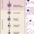
14 Nuclear and cytoplasmic changes in leukocytes
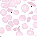
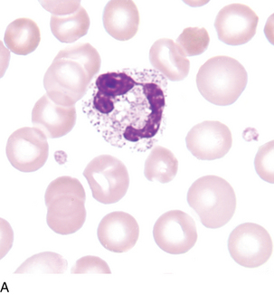
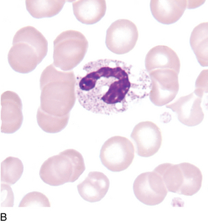
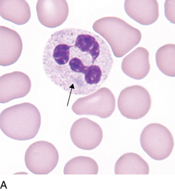
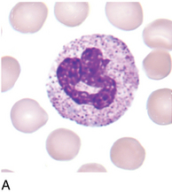
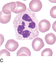
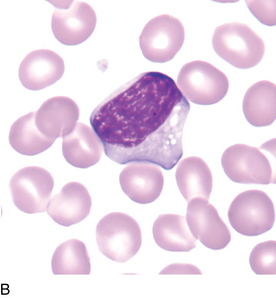
Hyposegmentation of neutrophils
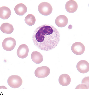
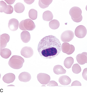
Description:
Associated with:
Pelger-Hüet anomaly, pseudo-Pelger-Hüet anomaly
Pelger-Hüet anomaly is inherited and affects the majority of granulocytes. Pseudo-Pelger–Hüet is acquired, affects less than 50% of granulocytes and is usually accompanied by other morphologic indications of malignancy such as seen in myeloproliferative or myelodysplastic disorders (see Chapters 17 and 18).
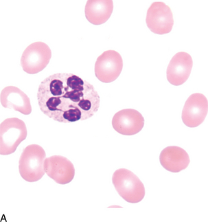
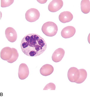
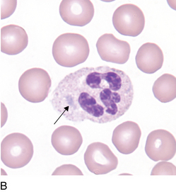
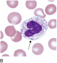

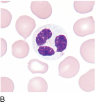
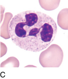
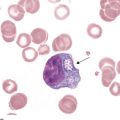
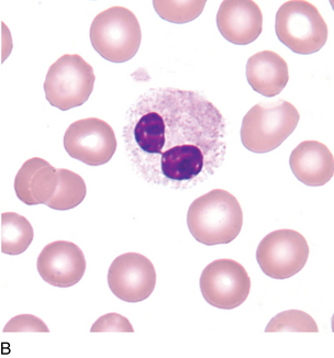
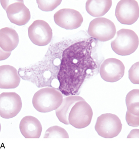
Reactive lymphocytes
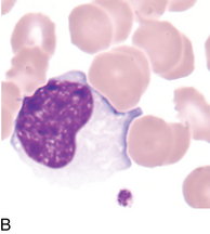
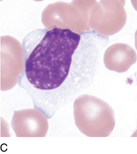
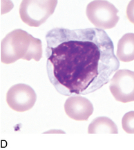
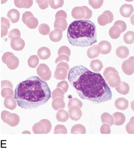
Figure 14–7E Reactive lymphocytes, characteristic of viral diseases, such as infectious mononucleosis (PB ×500).
Cytoplasm:
Pale blue to deeply basophilic, may stain unevenly with peripheral or radial basophilia
Associated with:
Viral infections and other antigenic stimulation, including organ transplantation
| Monocyte | Reactive Lymphocyte | |
|---|---|---|
| Shape | Pleomorphic; may have pseudopodia, which tend to “push away” surrounding cells | Pleomorphic, easily indented by surrounding cells |
| Size | 12-20 μm | 10-30 μm |
| Nucleus | Round, oval, horseshoe, or kidney shaped, may have brainlike convolutions | Irregular, elongated, stretched, occasionally round |
| Nucleoli | Absent | Occasionally present |
| Chromatin | Loosely woven, lacy | Variable; coarse to fine and dispersed |
| Cytoplasm | Blue-gray | Pale blue to deeply basophilic, may stain unevenly |
| Granules | Many fine red—may give ground glass appearance | May be a few prominent azurophilic granules |
| Vacuoles | Absent to numerous | Occasional |
Use as many criteria as possible to identify cells. It is often difficult to differentiate cells in isolation; multiple fields should be examined for nuclear and cytoplasmic characteristics. Consider “the company they keep.”
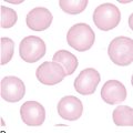
All photomicrographs are ×1000 with Wright-Giemsa stain unless stated otherwise.