Normal Retinal Anatomy and Basic Pathologic Appearances
Normal Retinal Anatomy
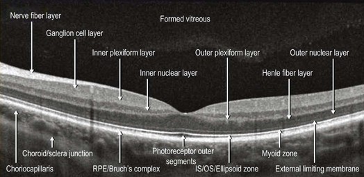
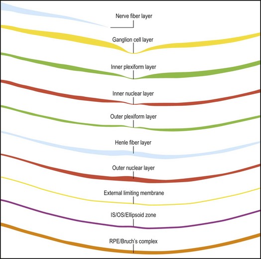
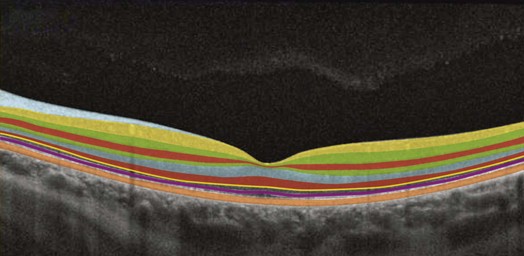
Commercially available SD-OCT scanners have an axial resolution of between 4 µm and 7 µm and a transverse resolution of approximately 15 µm. This high resolution allows for exquisite viewing of the retinal detail. Due to the limited penetration of light beyond the pigmented RPE and the drop-off of the OCT signal with depth, the image at the level of the choroid has lower resolution. The layers of the normal retina are labeled in Figure 4.1.1.
General Appearance of Retinal Pathology on SD-OCT
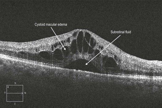
Cystic Changes in Outer Retina
Discrete hyporeflective spaces are noticed primarily in the outer retina, but usually span multiple layers (Fig. 4.1.3).
The differential diagnosis includes:
▶ Branch retinal vein occlusion
▶ Central retinal vein occlusion
▶ Retinal telangiectasias (e.g. Coat’s disease, macular telangiectasia)
▶ Vitreomacular disorders (vitreomacular traction, epiretinal membrane)
▶ Chronic subretinal fluid (e.g. retinal detachment, choroidal neovasclar membrane, central serous chorioretinopathy)
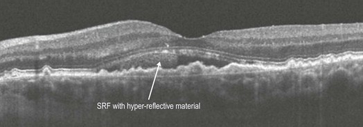
Subretinal Fluid
Clear hyporeflective space seen between the neurosensory retina and the RPE (Fig. 4.1.4).
The differential diagnosis includes:
▶ Central serous chorioretinopathy
▶ Choroidal neovascular membranes (secondary to e.g. age-related macular degeneration, myopia)
▶ Serous retinal detachments (secondary to tumors, inflammation, trauma)
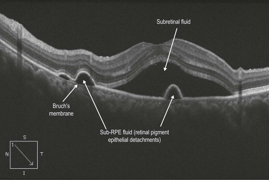
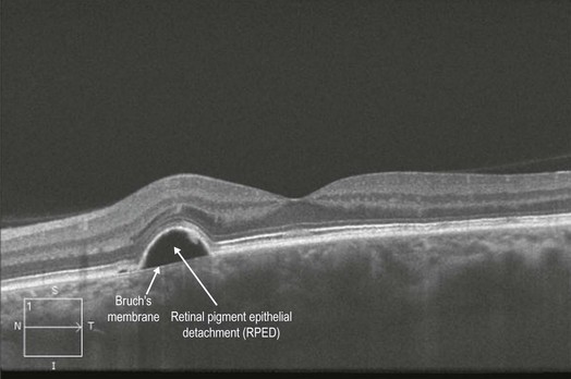
Retinal Pigment Epithelial Detachment
This is noted as a dome-shaped separation of the RPE from the underlying Bruch’s membrane. The ensuing space between the RPE and the Bruch’s membrane is hyporeflective (Fig. 4.1.6).
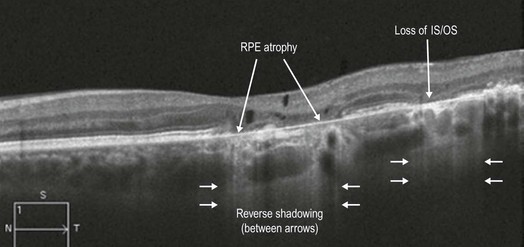
RPE Atrophy
Atrophy of the pigmented RPE causes decreased absorption of light. The OCT signal is therefore able to penetrate more deeply, which exaggerates the typical signal pattern so that there is a ‘reverse’ shadowing effect (Fig. 4.1.7, area between the smaller arrows)
The differential diagnosis includes:
▶ Geographic atrophy secondary to age-related macular degeneration
▶ Advanced chorioretinal scarring secondary to retinal degenerations and macular dytrophies (e.g. retinitis pigmentosa, Stargardt’s disease, cone dystrophy)
▶ Chorioretinal atrophy secondary to inflammatory disorders (e.g. ocular histoplasmosis, multifocal choroiditis)
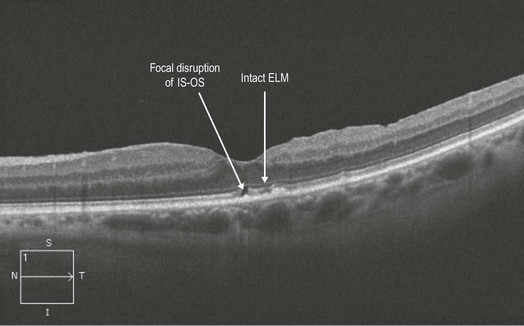
Focal Loss of External Limiting Membrane (ELM) and Inner Segment–Outer Segment (IS–OS) Photoreceptor Junction
OCT scanning reveals a disruption in the ELM line and in the IS–OS junction (Fig. 4.1.8). This is associated with a number of severe outer retinal conditions such as cone dystrophy and solar retinopathy as well as inner retinal disorders when they advance to involving the outer retinal layers. Loss of the IS–OS junction/ellipsoid layer as well as ELM has been associated with reduction in visual acuity and a worse prognosis for visual recovery in a number of ocular disorders.