Normal pregnancy and antenatal care

Published on 09/03/2015 by admin

Filed under Obstetrics & Gynecology

Last modified 22/04/2025

Print this page

rate 1 star rate 2 star rate 3 star rate 4 star rate 5 star
Your rating: none, Average: 0 (0 votes)

This article have been viewed 1731 times

Normal pregnancy and antenatal care

Shaylee Iles

Aims and patterns of routine antenatal care

The concept that the general wellbeing and reproductive performance of a woman might be improved by antenatal supervision is surprisingly recent, and was first introduced in Edinburgh in 1911. In many societies, antenatal care is not available or, for social or religious reasons, is not used when it is available. Unfortunately, it is often least available in those communities where the need is greatest and where antenatal disorders, particularly those linked to malnutrition or over nutrition, are most common.

The basic aims of antenatal care are:

The ways by which these objectives are achieved will vary according to the initial health and history of the mother and are a combination of screening tests, educational and emotional support and monitoring of fetal growth and maternal health throughout the pregnancy.

The frequency of antenatal visits was first established by a group of providers of antenatal care in 1929. The protocol advised that antenatal visits should occur monthly from 8 weeks gestation until 28 weeks and then every 2 weeks until 36 weeks and thereafter weekly until the time of delivery. In modern antenatal care, the timing of visits, particularly in the first 28 weeks of pregnancy, is now more closely geared to attendance for screening tests. In uncomplicated pregnancy, a reduction in the number of visits has not been shown to adversely affect maternal or perinatal outcome, although maternal satisfaction may be reduced.

Antenatal care is provided through a variety of different mechanisms and may be provided by general practitioners, midwives and obstetricians, often in a pattern of shared care. Pregnancies that are considered to be high risk should receive a high proportion of their care by obstetricians or specialists in fetomaternal medicine. Risk stratification should be assessed at the earliest antenatal visits and care planned accordingly. Guidelines for consultation and referral, such as those produced by the Australian College of Midwives or by the National Institute for Health and Clinical Excellence (NICE), can be a useful tool to assess risk and determine the most suitable model of care. Pregnancy risk and the most suitable care provider may alter during the course of pregnancy.

Preconceptual care and vitamin supplementation

Ideally, all women would present prior to conception to allow their health care professional to provide them with prepregnancy care and counselling. This role is often best provided by the woman’s usual general practitioner. This appointment allows for an opportunity to undertake screening tests and provide advice regarding conception and early pregnancy care.

Essential components of preconceptual care include the assessment of the need for immunization for rubella, varicella and pertussis. If the history of past vaccination or infection is uncertain, serology may be required. If serology is negative or immunization is due, this can then be provided. As these vaccines are live attenuated viral vaccines, it is recommended that the woman use adequate contraception to defer conception for 28 days following administration. Administration of the influenza vaccine on a seasonal basis to women who are intending to be or who are pregnant is also recommended due to the increased incidence of serious morbidity associated with influenza infection in pregnancy. This visit is also an ideal opportunity to undertake routine cervical cytology (Papanicolou smear) if due.

Dietary and vitamin supplementation advice should also be given at this time. It is recommended that all women take a folic acid supplement (400 µg daily) for at least one month prior to conception and the first three months of pregnancy as an effective means of reducing the incidence of neural tube defects. Certain risk groups may be recommended to take a higher dose (5 mg daily) such as those on anti-epileptic agents, obese women, diabetic women or women with a past history of neural tube defects. Iodine supplementation of 150 µg per day is also recommended in countries or regions where there is a dietary deficiency of iodine to aid in the development of the fetal brain. Some countries have overcome the problem by using iodized salts for cooking.

Maternal medical conditions, including medications, can be reviewed and optimized at this time. This provides an opportunity to discuss the impact of pregnancy on the medical condition as well as the impact of the medical condition on pregnancy. Medication may need to be altered or doses reduced where appropriate. Referral to specialist physician colleagues for treatment optimization may be appropriate.

Optimization of preconceptional health with advice on a nutritious diet and regular moderate exercise should also be provided at this time. Exploration and discussion around the use of licit and illicit substances should also be explored.

The risk of substance abuse in pregnancy

Smoking

Smoking has an adverse effect on fetal growth and development and is therefore contraindicated in pregnancy. The mechanisms for these effects are as follows (Fig. 7.1):

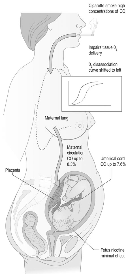
• The effect of carbon monoxide on the fetus. Carbon monoxide has an affinity for haemoglobin 200 times greater than oxygen. Fresh air contains up to 0.5 ppm of carbon monoxide, but in cigarette smoke values as high as 60 000 ppm may be detected. Carbon monoxide shifts the oxygen dissociation curve to the left in both fetal and maternal haemoglobin. Maternal carbon monoxide saturation may rise to 8% in the mother and 7% in the fetus, so that there is specific interference with oxygen transfer.

• The effect of nicotine on the uteroplacental vasculature as a vasoconstrictor. Animal studies on the effect of infusions of nicotine on cardiac output have shown that high-dose infusions produce a fall in cardiac output and uteroplacental blood flow. However, at levels up to five times greater than those seen in smokers there are no measurable effects and it is therefore unlikely that nicotine exerts any adverse effects by reducing uteroplacental blood flow.

• The effect of smoking on placental structure. Some changes are seen in the placental morphology. The trophoblastic basement membrane shows irregular thickening and some of the fetal capillaries show reduced calibre. These changes are not consistent or gross and are not associated with any gross reduction in placental size. The morphological changes have not been demonstrated in those women subjected to passive smoking.

• The effect on perinatal mortality. Smoking during pregnancy reduces the birth weight of the infant and also reduces the crown–heel length. Perinatal mortality is increased as a direct effect of smoking and this risk has been quantified at 20% for those women who smoke up to 20 cigarettes per day and 35% in excess of one packet per day. Mothers should be advised to stop smoking during pregnancy.

Alcohol intake

Excessive alcohol intake (in excess of eight standard drinks per day) is associated with a specific syndrome known as the fetal alcohol syndrome. Features in the infant include growth retardation, various structural defects and, in particular, facial defects, multiple joint anomalies and cardiac defects. However, these problems arise in women who consume 80 g of alcohol/day and who will almost inevitably have an unsatisfactory dietary intake as well. This is equivalent to an intake of 8 units/day, where 1 unit is equivalent to one glass of wine (200 mL) or half a pint of beer or lager. Increasingly, there is awareness of fetal alcohol spectrum disorder, a range of neurodevelopmental and behavioural effects attributable to alcohol consumption in pregnancy in a dose-dependent manner. Research is not clear as to what level of alcohol consumption is safe in pregnancy, so a recommendation to abstain from any consumption is the safest choice. In reality, the responsibility lies with the woman to adopt a reasonable approach to her alcohol intake. There is no evidence that the occasional social glass of wine or beer has any detrimental effect.

Illicit drug use

The common forms of drug abuse that occur during pregnancy are from heroin, amphetamines, cocaine and marijuana. All of these drugs have adverse effects on both the mother and the fetus, but many of the adverse effects are related to lifestyle and malnutrition.

Heroin addiction is associated with an increased incidence of intrauterine growth restriction, perinatal death and preterm labour. Furthermore, about 50% of infants exposed to heroin will suffer from neonatal withdrawal manifestations. The mother should be screened for HIV, syphilis, chlamydia and gonorrhoea and should be referred to a drug dependence unit for withdrawal of heroin and replacement with methadone or buprenorphine.

Amphetamine use has become an increasing problem over the past decade. Use in pregnancy is associated with an increased risk of miscarriage, preterm birth, growth restriction, placental abruption, fetal death in utero and developmental anomalies. Referral to a drug dependency service for advice on cessation is recommended.

Cocaine usage may induce cardiac arrhythmias and central nervous system damage in mothers as well as placental abruption, fetal growth restriction and preterm labour. Management of cocaine addiction is directed at withdrawing the drug.

Marijuana has no apparent adverse effect on pregnancy although the active ingredient of 9-tetrahydrocannabinol has been shown to have teratogenic effects in animal studies. Consumption is usually associated with significant tobacco use, which has major detrimental effects as outlined above.

Changing demographics of pregnancy

Maternal age is an important determinant of outcome in obstetric services, with increased risk being associated with both extremes of maternal age. In recent years the median age of women giving birth in developed countries has continued to rise, and currently sits at around 30 years. The reasons for this are complex and due to a number of social, economic, and educational factors. Rates of pregnancy in the over 35 age group continue to rise (currently 23% of all births) as does that of women over 40 (4% of all births), however the number of mothers over 45 years remains low.

Access to assisted reproductive technologies (ART) has increased with a subsequent influence on median maternal age. Approximately 3.2% of babies born are conceptions assisted by ART in Australia and the UK. In addition, rates of multiple pregnancies continue to rise, currently around 1.6% of all mothers. This is largely due to the increases in ART and increasing maternal age. Rates of multiples in ART pregnancy are around 10% of all successful conceptions.

Rates of teenage pregnancy continue to decline and sit at around 4% of all births.

The absolute number of babies born to each woman continues to be low, with 75% of mothers giving birth to their first or second baby. The median age of first time mothers also continues to climb, and is currently around 28 years.

Women are active participants in antenatal care with over 98% having at least one antenatal visit and 92% having 5 or more visits. Preterm birth occurs in around 7.5% of all pregnancies, with most of these occurring in gestations greater than 32 weeks.

Around 75% of all women use analgesia in labour, most commonly nitrous oxide, followed by opioids, then regional techniques (predominantly epidural anaesthesia, around 30%). Rates in first time mothers of analgesic use are around 85%. Rates of caesarean section as a proportion of births increase with increasing maternal age.

The booking visit

The details of antenatal history and routine clinical examinations are discussed in Chapter 6. However, certain observations should be stressed at the first visit and it is preferable that these observations should be made within the first 10 weeks of pregnancy. The measurement of maternal height and weight is important and has value in prediction of pregnancy outcomes. Women with a low body mass index (BMI; less than 20, where BMI is estimated as weight (kg) divided by height (m2)) are at increased risk of fetal growth restriction and perinatal mortality. Women with a high BMI are increasingly recognized as being at increased antenatal and intrapartum risk, with the risks beginning to rise from a BMI of 30.

The initial measurement of blood pressure should be taken as soon as possible as this may provide evidence that, if there is hypertension, it is likely to have predated the pregnancy.

Consideration of past obstetric history, including mode of delivery

A record should be made of all previous pregnancies, including previous miscarriages and terminations, and the duration of gestation in each pregnancy. In particular, it is important to note any previous antenatal complications, details of induction of labour, the duration of labour, the presentation and the method of delivery, as well as the birth weight and gender of each infant. The mode of delivery (spontaneous, assisted or caesarean section) has implications for the current birth and needs to be explored. Previous operation records should be sought if relevant to aid in appropriate counselling for this pregnancy.

The condition of each infant at birth and the need for care in a special care baby unit should be noted.

Complications of the puerperium such as postpartum haemorrhage, extensive perineal trauma or wound breakdown, infections of the genital tract, deep vein thrombosis or difficulties with breastfeeding may all be relevant to the current pregnancy.

Recommended routine screening tests

Beginning at the first visit, a number of screening tests are introduced. Some will be repeated later in the pregnancy. The omission of these tests will generally now be considered to be evidence of substandard practice so they have medicolegal importance as well as clinical relevance.

Haematological investigations

Anaemia is a common disorder in pregnancy and in most communities will be due to iron deficiency, either because of the depletion of iron stores or because of reduced iron intake. Over 90% of pathological anaemia in pregnancy is due to iron deficiency. However, it may also be macrocytic and due to folate or vitamin B12 deficiency or may be related to various parasitic infections.

Haemoglobin concentration and a full blood count should therefore be performed at the first visit and repeated at 28 and 34 weeks gestation. Women who have deficient iron intakes should be given oral supplements of iron from early in pregnancy. Screening for haemoglobinopathies should be routinely offered to those racial groups where conditions such as thalassaemia and sickle cell disease are common.

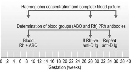
Blood group and antibodies

Blood group should be determined in all pregnant women and screening for red cell antibodies should be undertaken early in pregnancy. In Rhesus (Rh)-negative women, screening for Rh antibodies should be performed at the first visit (preferably in the first trimester) and then repeated at least at 28 weeks gestation. ABO antibodies may also cause problems in the fetus and newborn, but there is no method available to counter this problem.

The use of anti-D immunoglobulin

Around 15% of Caucasian women will be Rh negative and be at risk of developing anti-D antibodies during or immediately following pregnancy. The formation of anti-D antibodies may pose a risk to the wellbeing and even survival of a subsequent fetus due to the preformed antibodies crossing the placenta and attacking the red blood cells of a Rh-positive fetus. The effects on the fetus and newborn can be devastating and include anaemia, hydrops, neonatal anaemia, jaundice, kernicterus or fetal death in utero. There is very strong evidence dating from the 1960s that postpartum administration of anti-D immunoglobulin (anti-D Ig) can dramatically reduce the incidence of this complication.

Until the past few years, anti-D Ig was given only to women with a sensitizing event in pregnancy or postnatally to women delivered of a Rh-positive infant. Given within 72 hours of birth, this dose reduces the risk of Rh isoimmunization to around 1.5%. Quantitation of the degree of fetomaternal haemorrhage and the need for further doses should be undertaken by flow cytometry (where available) or the Kleihauer-Betke test prior to administration of the first dose.

Sensitizing events include normal delivery, miscarriage, termination of pregnancy, ectopic pregnancy, invasive prenatal diagnosis, abdominal trauma, antepartum haemorrhage or external cephalic version.

Now that anti-D Ig is readily available, it has become standard practice to give anti-D Ig prophylaxis at 28 and 34 weeks gestation (Fig. 7.2). This will prevent maternal immunization by a Rh-positive fetus in all but 0.2% Rh-negative women, in whom the infusion of cells from the fetus overwhelms the dose of antibody administered. This is in addition to the above indications.

Infection screening

Rubella

All females are offered rubella vaccination between the ages of 11 and 14 years, often through a school-based vaccination programme. By the time they present for their first confinement, 22% of nulliparous women will still be found to be non-immune, as well as 1.2% of multiparous women. Around 50% of non-immune women will have been previously vaccinated. All seronegative women should be offered immunization in the immediate puerperium. Vaccination is performed with a live attenuated rubella virus vaccine and involves a single dose injected subcutaneously. Although there is no evidence to suggest any significant increase in abnormality rate in the babies in women who have conceived immediately before or following rubella vaccination, it is generally recommended that pregnancy should be avoided for 1 month after vaccination. Non-immune women should be advised to avoid contact with infected individuals. Any clinically suspected infection should be investigated with paired sera, preferably with the original sample taken at the time of booking.

Syphilis

Routine screening for syphilis is recommended practice. Despite the fact that the condition is now relatively rare, the condition is treatable and has major neonatal sequelae if left untreated. Various tests are available.

Non-specific tests

The Wasserman reaction is a complement-fixation test that was the first successful serological test described for use in clinical practice. The test is dependent on the presence of treponemal antibodies in the serum, which unite with a colloidal suspension of lipoids to produce visible flocculation. A similar flocculation test that is widely used is the venereal disease research laboratory test (VDRL), which employs a cardiolipin antigen. The rapid plasma reagin (RPR) test is commonly used as it is an inexpensive test and is used primarily for screening and for follow up to check if there is a response to treatment. The difficulty with these tests is that they may give a false-positive reaction in association with malaria or viral pneumonia, or in autoimmune conditions such as lupus erythematosus, haemolytic anaemias, Hashimoto’s disease or rheumatoid arthritis. The VDRL test usually becomes negative within 6 months of treatment and therefore has an important role in treatment monitoring.

Human immunodeficiency virus

The basis of tests for the detection of human immunodeficiency virus (HIV) is the detection of HIV antibodies. The virus can be isolated and grown but this is a difficult procedure. As the virus has a predilection for the T-helper subset of lymphocytes, there is an altered T-helper/T-suppressor ratio. However, all these tests can be normal, even in the presence of infection. The most important confounding variable is that HIV antibodies may be absent in the incubation phase.

Seropositive mothers always have seropositive babies due to transplacental transmission of antibodies, but this may not indicate active infection in the baby. However, up to 45% of babies will have contracted HIV if active management programmes are not used. As treatment is highly effective in reducing transmission rates to less than 2% there is a strong case for routine screening of all women. These strategies include caesarean section, avoidance of breastfeeding and antiretroviral therapy in both the antenatal and intrapartum period as well as for the newborn (see Chapter 9).

Group B Streptococcus

Group B Streptococcus (GBS) is a Gram-positive bacterium that is a common commensal carried in the gastrointestinal tract. It can be cultured from the vagina in up to 25% of women in pregnancy and may also be a cause of urinary tract infection. During vaginal delivery there is a risk of transmission to the neonate. This risk is increased in preterm delivery and prolonged rupture of membranes. Neonatal infection occurs in 1–2 per 1000 births and can result in overwhelming sepsis associated with significant morbidity and mortality. Ninety percent of infections present within the first days of life, but late presentations at up to 3 months of age can occur.

The organism can be detected on vaginal and rectal swabs and the rate of vertical transmission reduced by the use of intrapartum antibiotic treatment with intravenous penicillin. Screening for GBS using a low vaginal swab taken between 34 and 36 weeks is recommended by many centres but is not universal practice.

Gestational diabetes

Gestational diabetes is associated with an increased incidence of intrauterine fetal death as well as intrapartum and neonatal complications. Screening programmes follow one of two pathways:

• Selection by history:

Under these circumstances, a full glucose tolerance test (GTT) should be performed using either a 75 g or 100 g loading dose of glucose. The test should be performed at the booking visit and again at 28 weeks gestation if there is any doubt about the diagnosis.

• Universal screening: The screening of all women at 26–28 weeks gestation will identify more women with impaired glucose tolerance or diabetes than those screened by risk factors alone. A modified GTT involving a loading dose of 50 g and 1 hour blood glucose (glucose challenge test, GCT) is considered positive if the blood glucose exceeds 7.7 mmol/L. This is then followed by a formal GTT.

Most units prefer to screen at-risk populations because of the practical difficulties and costs of screening the whole population, particularly in large maternity hospitals.

Screening for fetal anomaly

Structural fetal anomalies account for some 20–25% of all perinatal deaths and for about 15% of all deaths in the first year of life. There is therefore a strong case to be made for early detection and termination of pregnancy offered where this is appropriate. The frequency of the major structural anomalies is shown in Table 7.1. Congenital anomalies are one of the markers of socioeconomic deprivation.

Table 7.1

Structural anomalies

Type of anomaly Frequency (per 1000)
Cardiovascular 6
Craniospinal 3–7
Renal tract 1
Gastrointestinal 1

These anomalies are generally detectable by ultrasound scanning and this will be discussed in Chapter 10.

Nuchal translucency and biochemical screening

Screening for trisomy 21 (Down’s syndrome) has become routine in most antenatal services, but not in all countries. The logical consequence of such a programme is to offer invasive testing then termination of pregnancy where there is evidence of aneuploidy. Although the value of the test is reduced if termination is not an option, a positive result can help parents prepare for the birth of an affected infant. Screening is by the use of biochemical and ultrasound tests. It is important that women understand that these are screening tests and therefore have their limitations. They will not detect every case and high-risk results do not necessarily mean that the baby is affected. Despite the increased incidence of Down’s syndrome in mothers over 35 years, screening on the basis of age alone will not detect most affected fetuses and it is recommended to offer screening to all women. The major modality for screening for Down’s syndrome is the use of ultrasound measurement of nuchal translucency, a measurement of fluid behind the fetal neck (see Chapter 10, Fig. 10.6). This is combined with maternal age and the results of biochemical tests to provide a risk for this fetus of trisomy 21, 13 and 18 (see Chapter 10).

Schedules of routine antenatal care

Subsequent visits

Although the pattern of antenatal care will vary with circumstances and with the normality or otherwise of the pregnancy, a general pattern of visits will partly revolve around the demands of the screening procedures and the obstetric and medical history of the mother. The measurement of blood pressure is performed at all visits and the measurement of symphysis/fundal height should be recorded, even accepting that this observation has a limited capacity to detect fetal growth restriction. Serial ultrasound measurements would have a greater detection rate if performed at every visit, but this is not practicable or necessary for women who are not considered to be at high risk. A suggested regime for antenatal visits is listed in Table 7.2.

Table 7.2

Visits for antenatal care

8–12 weeks Initial visit, confirmation of pregnancy, search for risk factors in maternal history. Cervical smear where indicated, advice on general health, smoking and diet. Discuss and organize screening procedures. Check maternal weight and give advice on recommended folic acid and iodine supplements. Dating scan and scan for multiple pregnancies
11–14 weeks Screening for trisomies with ± nuchal translucency scan and blood tests if requested. Confirm booking arrangements. Offer dietary supplements of iron if any evidence of anaemia
16 weeks Check all blood results. Offer routine ultrasound anomaly scan
20 weeks Check ultrasound result. BP. Fundal height.
24 weeks BP, fundal height, fetal activity.
28 weeks BP, fundal height, fetal activity, full blood count and antibody screen. Administer anti-D if Rh-negative. Glucose tolerance test.
32 weeks BP, fundal height, fetal activity and fetal growth scan where pattern of fetal growth is in doubt or low lying placenta on anomaly scan
34 weeks Routine checks, also second dose of anti-D for Rh-negative women, Group B Streptococcus vaginal and rectal swab, full blood count
36 weeks BP, fundal height, fetal activity, determine presentation
38 weeks Routine checks, fetal activity, maternal wellbeing
40 weeks Routine checks, fetal activity, maternal wellbeing
41 weeks Routine checks, assessment by pelvic examination as to cervical favourability, cardiotocograph, amniotic fluid index. Individualize care with regards to induction of labour and ongoing assessment.

(Adapted from Kean L (2001) Routine antenatal management. Curr Obstet Gynaecol 11:63–69.)

In general, where pregnancies have been accurately dated by early ultrasound so that the gestational age is certain, induction of labour after 41 weeks reduces the incidence of meconium staining, macrosomia and the risk of fetal and neonatal death. Although the meta-analysis suggests there is reduction of caesarean and instrumental deliveries with induction of labour, this has been challenged, as the methods used for induction in the conservative group in the largest study did not use prostaglandin for ripening or induction of labour.

Antenatal education

An important and integral part of antenatal care is the education of the mother and her partner about pregnancy, childbirth and the care of the infant. This process should start before pregnancy as part of school education and should continue throughout pregnancy and the puerperium. There are various ways by which this can be achieved but, commonly, the needs are met by regular antenatal classes during the course of the pregnancy. It is preferable that those staff who are involved in general antenatal care and delivery should be part of the team that delivers the woman so that the processes of care and education are seen as one entity.

Dietary advice

There can be no doubt about the importance of diet in pregnancy. At one extreme, gross malnutrition is known to result in intrauterine growth retardation, anaemia, prematurity and fetal malformation. Lesser degrees of malnutrition may also be associated with an increased incidence of fetal malformations, particularly neural tube defects, and it is therefore important to provide guidance on diet and to ensure that a diet of appropriate quality and quantity is maintained throughout pregnancy and the puerperium.

Clearly, there will be substantial variation in the nature of the diet depending on racial group and actual physical size, but there are general principles that can be laid down as advice to meet the needs of the mother and of the developing fetus.

Advice should be given early in pregnancy regarding Listeria risk and advice given to avoid high-risk foods such as soft cheeses, delicatessen meats, salad bars and soft serve ice cream.

Minerals and vitamins

Other than folic acid and possibly iodine, routine supplementation with iron and vitamins should not be necessary during pregnancy. However, where there is evidence of dietary deficiency or in cases of multiple gestations, iron and vitamin supplements should be given from the first trimester onwards.

The requirements for iron, calcium, iodine and various trace elements such as magnesium and zinc are all increased in pregnancy. These elements are found in lean meat, various stone fruits, beans and peas, dairy produce and seafood.

Vitamins A and B are found in kidney, liver and dark green vegetables. Vitamin B2 is found in whole grain and cereals, and Vitamin B5 in fish, lean meat, poultry and nuts.

Ascorbic acid is essential for fetal growth and maternal health and is found in citrus fruits, brussels sprouts and broccoli. Vitamin D and folic acid are also important. Vitamin D deficiency in pregnancy is becoming increasingly common. In those women who cover themselves completely for religious reasons or for protection against skin cancers, there is a risk of vitamin D deficiency. Testing and supplementation as needed from early pregnancy is recommended. Folic acid deficiency is still relatively common and is associated with the development of megaloblastic anaemia in pregnancy. Green vegetables, nuts and yeast are all rich sources of folic acid and are available and inexpensive year round in supermarkets so there is no need for these deficiencies to arise.

A general protocol for diet in pregnancy is given in Table 7.3.

Table 7.3

General advice on foodstuffs recommended in pregnancy (quantity per day unless otherwise stated)

Foodstuff Quantity
Dairy Milk 600–1000 mL
Butter 150 g
Cheese 1 serving
Meat Chicken, pork or beef 2 servings
Liver Once a week or more
Fish Once or twice a week
Vegetables Potato 1–2 servings
Other 1–2 servings
Salads Freely
Fruit Citrus 1 serving
Other 2–3
Cereals Wheat, maize, rice, pasta 4 servings

image

1 serving = half a cup.

Breast care

Breastfeeding should be encouraged in all women unless there are specific contraindications that would have adverse fetal or maternal consequences. Previous damage to the breasts or grossly inverted nipples may make breast feeding difficult. There are also medications that are concentrated in breast milk and may be hazardous for the infant, in which case breastfeeding is contraindicated. In some maternal infections, such as HIV, breastfeeding is contraindicated. However, these circumstances are uncommon and, in most conditions, the mother should be advised of the benefits to both her child and herself of breast milk.

In the antenatal period, good personal hygiene including breast care should be encouraged. Colostrum may leak from the nipples, particularly in the third trimester, especially in multiparous women. The breasts should be supported with an appropriate maternity brassière. Antenatal referral to a lactation consultant for women who have risk factors for potentially encountering difficulty with breastfeeding such as previous difficulty, or breast surgery, should be offered.

Safe prescribing in pregnancy

The use of prescription and over the counter medications, as well as complementary and alternative medications, is common. Some women will require ongoing treatment of pre-existing medical conditions, e.g. epilepsy or asthma. Some conditions may develop de-novo in pregnancies that require therapy, e.g. gestational diabetes, thromboembolism. Simple analgesics, antipyretics, antihistamines and anti-emetics are all commonly consumed. A discussion of the risks and benefits of individual medications is beyond the scope of this text. Extensive information is available in most drug formularies about the safety of categories of drugs in pregnancy and lactation. Reputable online resources such as www.motherisk.org are available around the clock and often helpful. The safest course before prescribing in pregnancy is to always check. Many medications have been shown to have no adverse outcomes when used in pregnancy or lactation.

In general, simple analgesia is best provided by paracetamol which remains a safe drug to consume in pregnancy. Non-steroidal anti-inflammatory drugs are generally contraindicated due to fetal effects. Metoclopramide as a first line anti-emetic is safe to consume in pregnancy, including during embryogenesis in the first trimester.