23 Normal newborn peripheral blood morphology
In the healthy, full-term newborn, peripheral blood collected within the first 12 hours of birth has distinctive morphology. Some morphological changes persist for up to 3 to 5 days after birth. These changes should be recognized as physiological and not pathological. For a fuller discussion of hematology in the newborn, refer to a hematology textbook such as Hematology: Clinical Principles and Applications* or a pediatric hematology text such as Nathan and Oski’s Hematology of Infancy and Childhood.†
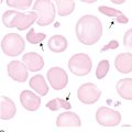
Occasional spherocytes are common, varying from one every two fields to one or more in every field.
Newborn total leukocyte counts are higher than for adults, and newborns have more segmented and band neutrophils than at any other time in childhood.‡ An occasional metamyelocyte may be seen without evidence of infection. Monocyte morphology is similar to that of the adult.
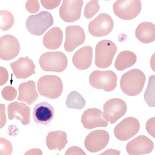
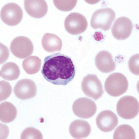
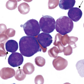
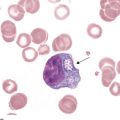
FIGURE 23–4 Bone marrow from neonate with acute lymphoblastic leukemia, demonstrating hematogones and lymphoblasts. Hematogones vary in size. Nucleus is round to oval with condensed, smudged chromatin. Nucleoli are absent or indistinct. Cytoplasm is indiscernible to scant. Arrows point to hematogones. Most of the other cells are blasts (BM ×1000). See chapter 16 for comparison with blasts.
* Rodak BF, Fritsma GA, Keohane EM, editors: Hematology: clinical principles and applications, ed 4, St. Louis, 2012, Saunders.
† Orkin SH, Nathan DG, Ginsburg D et al: Nathan and Oski’s hematology of infancy and childhood, ed 7, St. Louis, 2009, Saunders.
‡ Quinn CT, Buchanan GR: Hematopoiesis and hematologic diseases. In McMillan JA, Feigin RD, DeAngelis C, Jones MD, editors: Oski’s pediatrics, Philadelphia, 2006, Lippincott Williams & Wilkins.