Lymph Nodes, Enlarged
Synonyms/Description
Etiology
Ultrasound Findings
Differential Diagnosis
Clinical Aspects and Recommendations
Figures
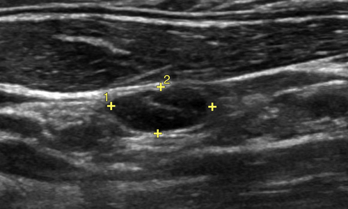
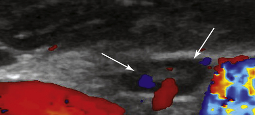
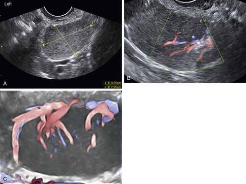
Figure L1-3 A, Magnified view of a left-sided mass seen transvaginally, showing a homogeneously solid mass (calipers). This enlarged and abnormal node is rounded and wide, with loss of normal architecture. B, The same mass is interrogated with color flow Doppler, showing that the blood flow into the mass originates from one side, characteristic of a lymph node. C, The contralateral lymph node (seen here using 3-D color Doppler) is also enlarged with abundant color flow.
Suggested Reading
Fischerova D. Ultrasound scanning of the pelvis and abdomen for staging of gynecological tumors: a review. Ultrasound Obstet Gynecol. 2011;38:246–266.
Lai G., Rockall A.G. Lymph node imaging in gynecologic malignancy. Semin Ultrasound CT MRI. 2010;31:363–376.
Gore R.M., Newmark G.M., Thakrar K.H., Mehta U.K., Berlin J.W. Pelvic incidentalomas. Cancer Imaging. 2010;10:S15–S26.


