120
Nevus sebaceus
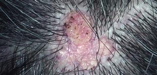
Nevus sebaceus in a 30-year-old man. The plaque has developed in to a large verrucous yellow-pink nodule with a decrease in terminal scalp hair.
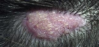
This 12-year-old boy has reached puberty, and the lesion has become thicker. This change at puberty is expected and represents, in part, sebaceous gland and epithelial hyperplasia.
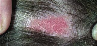
Nevus sebaceus occurs most often on the scalp, although lesions are also found on the face, neck, and trunk.
The lesion is usually a solitary round to oval plaque but it may be linear and disjointed.
DESCRIPTION
Distinctive congenital lesion of the head (usually scalp), composed of skin and appendageal components. Nearly all nevus sebaceus lesions are present at birth or appear in early childhood. Lesions change clinically and histologically with age. Not all lesions may be noticed at birth. At puberty, these nevi tend to enlarge and may be noticed for the first time at this point.
HISTORY
• Nevus sebaceus remains stable throughout childhood and undergoes predictable change at puberty. • In the first few months of life, the sebaceous glands are well developed as a result of maternal hormonal stimulation, although surrounding hair structures are incompletely differentiated. Thereafter and through the rest of childhood, the sebaceous glands are small in size and number; incompletely developed hair structures may be seen. • With puberty, hormonal influences bring about diagnostic changes. Sebaceous glands mature and increase in size and density. Hair structures remain undifferentiated, and papillomatous epidermal hyperplasia develops. Ectopic apocrine glands may also be found deep within the underlying dermis. • Appendageal tumors may develop later in life within nevus sebaceus. Each such tumor has its own histologic pattern. The most common tumor is syringocystadenoma papilliferum, a benign apocrine tumor seen in up to 20% of lesions. Basal cell carcinoma is the second most common tumor and most common malignancy that develops in nevus sebaceus. It occurs in roughly 7% of lesions, but rarely, if ever, metastasizes.
PHYSICAL FINDINGS
Nevus sebaceus is usually a solitary, yellowish to flesh-colored plaque and occurs most commonly on the scalp, forehead, or postauricular areas. It tends to be linear or oval-shaped, measuring 1–3 cm in diameter. The lesion evolves in three stages corresponding to sebaceous gland maturation through childhood, puberty, and adulthood. • In childhood, the plaque is barely raised and has a velvety surface, is hairless, pink to tan, and asymptomatic. • Around puberty, the plaque tends to thicken, become larger and more verrucous, and has a yellow-white and pink-speckled appearance. Lesions at this stage are easily traumatized and may be tender. • The third stage of evolution occurs during adulthood. Nevus sebaceus may be confused with linear epidermal nevus clinically.
TREATMENT
• Excision of the entire lesion is recommended; nevus sebaceus has a tendency to develop basal cell carcinoma within the nevus after puberty. • Excision is best performed just before puberty, when the lesion is still small and the patient is old enough to understand and tolerate the procedure. • A larger lesion may be excised in stages without any concern of inducing malignant change.


