135
Nevi, melanocytic nevi, moles
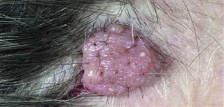
This is a common presentation for a scalp nevus. There is no pigment, and the surface is lobulated.
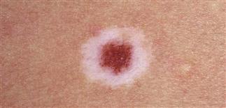
Halo nevus. A white halo appeared around a benign-appearing nevus. The nevus then slowly disappeared over a 3 year period.
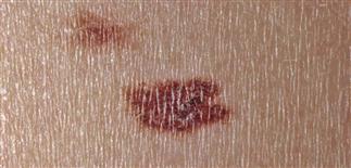
A junction nevus with a slightly raised, brown, uniform surface. The lesion had been present for years.
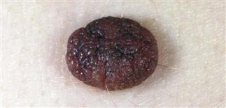
A dermal nevus with horn pearls on the surface. It is stuck on to the surface and has the consistency of a seborrheic keratosis.
DESCRIPTION
Benign growths composed of melanocyte-derived nevus cells, classified by age of onset, arrangement of nevus cells within skin.
HISTORY
• Nevi are ubiquitous; most adults have 12–20. • Incidence of acquired nevi peaks during adolescence. Few appear after age 30. • Most acquired nevi appear on sun-exposed skin, are asymptomatic. Consider nevi in sun-protected areas suspicious. • May become darker during puberty, during pregnancy. • Acquired nevi begin as flat, round, uniformly colored papules. Nevi mature by expanding laterally and symmetrically. • Usually elevate and lighten over time, eventually becoming a skin-colored papule.
PHYSICAL FINDINGS
• Junctional nevi. Flat or slightly raised brown to tan macules, mostly in children. Nevus cells cluster at dermoepidermal junction. Nevi of palms, soles, genitalia, mucosa usually junctional. • Compound nevi. Raised pigmented papules. Nevus cells found at dermoepidermal junction and within dermis. Can have irregular border but are symmetric. • Intradermal nevi. Usually elevated, fleshy, pigmented papules, but may contain no pigment. Nevus cells found within dermis, sometimes extending into subcutaneous fat. • Nevus spilus. Similar to a café-au-lait spot but contains small, monomorphic, dark brown junctional nevi. • Blue nevi. Solitary, dark bluish papules usually on head, neck, buttocks. Color due to intensely pigmented melanocytes in deep dermis. • Spitz nevus. Reddish pink, dome-shaped, smooth papule found usually on face, scalp, limbs of children. While benign, Spitz nevi contain pleomorphic nevus cells. Most dermatologists favor complete removal. • Halo nevi. Occur primarily during adolescence. A pre-existing nevus develops surrounding hypopigmentation then gradual disappearance of nevus. Halo nevi appear to be a host response directed against nevus cells. • Recurrent nevus phenomenon. May occur at site of previously partially removed nevus. Random pleomorphic nevus cells along with scar can be suspicious for melanoma.
Most melanocytic nevi are benign and follow the course of maturation described above. Nevi that deviate from this pattern are suspicious and biopsy is warranted.
TREATMENT
• Assess all nevi regularly, carefully, singly and in aggregate. • Most benign nevi symmetric, less than 6 mm in diameter, with well-defined, regular border, uniform color. • Regard nevi appearing different from others on same patient with suspicion. • Biopsy suspicious nevi. • Examine entire cutaneous surface. • Educate patients to self-examine all skin areas periodically. Review changes to watch for.


