CHAPTER 18 Neurosurgery
Head injuries
 It is recommended that all patients are monitored using the Glasgow Coma Scale after initial resuscitation (→ Table 4.1, in Ch. 4). In all cases, the diagnosis and initial treatment of serious extracranial injuries takes priority over investigations of head injury, or transfer to a neurosurgical unit.
Criteria for urgent CT scan and consultation with a neurosurgical unit
The presence of one or more of the following:
Criteria for hospital admission after recent head injury

 Simple scalp laceration is not a criterion for skull radiography if the history can exclude a significant impact.
Assessment of head injury
Emergency

Evaluate CNS injury
Assess the level of consciousness as this is the most significant factor after head injury. Use GCS (→ Table 4.1) and check pupillary reactions. Pupillary changes may indicate brain swelling or compression. Pressure on a cerebral hemisphere causes the third nerve on that side to be stretched over the edge of the tentorium. The resultant paralysis of the nerve allows unopposed action of the dilator pupillae under the control of the sympathetic nervous system and the pupils dilate. There is also loss of light reaction of the pupil on the affected side. If compression continues, the contralateral third nerve is compressed and the opposite pupil also dilates and is fixed to light. Bilateral fixed dilated pupils in a patient with a head injury are a grave prognostic sign and recovery is rare. Pupillary changes are always late signs (‘undertaker signs’), and are always preceded by an alteration in conscious level caused by raised intracranial pressure. Direct blows to the eyes can cause dilated pupils in patients without severe brain injury.
Check for CSF rhinorrhoea or otorrhoea
Check for these signs. Periorbital bruising and retromastoid bruising imply basal skull fracture.
Scalp lacerations or depressed fractures
Check these using a gloved finger. If in doubt, perform a CT scan.
Skull fractures
They may be further classified as follows:
Intracranial bleeding
Management of head injuries
 Always consider – is this only a head injury or was it caused by something else, e.g. myocardial infarction, fit, intracranial bleed? A good history is vital.
Minor
Observations during admission
The primary observations are the GCS (→ Table 4.1). In addition pulse, BP, respiratory rate and pupillary size and reaction are monitored. These are carried out by the nursing staff on the ward, the frequency depending on the severity of the symptoms. Hourly observations are usually the norm. Signs of deterioration include falling coma score, falling pulse rate, raised BP, reduced or irregular respirations, dilatation of the pupils, loss of light reflex and asymmetrical pupils. An alteration of conscious level occurs before signs of brainstem compression.
Other complications of head injury
Brain death
There are prerequisites for the diagnosis of brainstem death:
Management of raised intracranial pressure (ICP)
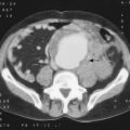
 Causes Head injury, meningoencephalitis, haemorrhage (extradural, subdural, subarachnoid, intracerebral), tumour, infection, hydrocephalus.
 Lumbar puncture should not be carried out in the presence of raised ICP. If the spinal CSF pressure is reduced by removing CSF, the high ICP may force the brainstem and cerebellar tonsils through the foramen magnum (coning) with fatal results.
Cerebral tumours
These may be broadly classified as glial and non-glial depending on the cell of origin (→ Table 18.1).
| Primary | |
| Glial (gliomas) | Astrocytoma |
| Medulloblastoma | |
| Ependymomas | |
| Oligodendrogliomas | |
| Non-glial | Meningiomas |
| Acoustic neuroma | |
| Pituitary tumours | |
| Secondary | Lung |
| Breast | |
| Kidney | |
| Melanoma |
Pituitary tumours
Intracranial vascular lesions
Hydrocephalus
Clinically, hydrocephalus may be divided into two groups, congenital and acquired.
Spinal tumours
| Extradural | Secondary spinal deposits are most common |
| Primary bone tumours, e.g. osteoblastoma and myeloma | |
| Intradural–extramedullary | Meningioma |
| Neurofibroma | |
| Intramedullary | Rare and include astrocytomas and ependymomas |