Neurologic Aspects of Chronic Pain

Published on 03/03/2015 by admin

Filed under Neurology

Last modified 22/04/2025

Print this page

rate 1 star rate 2 star rate 3 star rate 4 star rate 5 star
Your rating: none, Average: 0 (0 votes)

This article have been viewed 1434 times

Chapter 14 Neurologic Aspects of Chronic Pain

More than a persistent symptom, chronic pain is a complex endless condition with unique qualities, impaired activities of daily living, and psychiatric comorbidities. Traditional medical approaches to chronic pain emphasized diagnosis, distinguished between its psychologic and physiologic components, and sought its eradication. Currently, multidisciplinary pain teams stress symptomatic management, reducing pain’s affective component (suffering), and restoring function. These teams describe their role as pain management specialists without holding out the expectation that they will eliminate the pain.

As a member of a team or an individual-treating physician, psychiatrists should be aware of common chronic pain syndromes and their underlying neuroanatomy, psychiatric comorbidity, and treatments involving long-term use of opioids (narcotics) and adjuvant medications (primarily nonanalgesic drugs, such as antidepressants and antiepileptic drugs [AEDs]).

Pain Varieties

Nociceptive pain results from acute, ongoing tissue damage, such as a metastasis to bone, dental infection, or disease of the viscera. It usually consists of dull aching pain at the site of the tissue damage. Diseased viscera, however, may refer pain to another region, such as when gallbladder stones seem to produce pain in the scapula. Wherever the location, tissue damage triggers specific receptors (nociceptors) in the peripheral nervous system (PNS) and certain cranial nerves. Either peripheral or cranial nerves transmit the noxious stimuli to the central nervous system (CNS). Removing diseased tissue and other direct treatments reduce or eliminate nociceptive pain. Until the injury responds to treatment or heals, analgesics reduce the pain.

Neuropathic pain, in contrast, results from direct nerve tissue injury. It is a symptom of a wide variety of CNS and PNS disorders, such as cranial neuropathies, mononeuropathy or polyneuropathy, brachial or lumbar plexus injury, lumbar spine disorders, complex regional pain syndrome, and thalamic injury.

Unlike the dull ache of nociceptive pain, neuropathic pain consists of electric, sharp, lancinating, or burning sensations. Also, not confined to the site of tissue injury, neuropathic pain and spontaneously occurring painful paresthesias radiate throughout the distribution of the injured nerve and often well beyond it. Other features are that painful or even neutral stimuli elicit an intense, distorted, or prolonged response – allodynia, hyperalgesia, and hyperpathia (see Chapter 5).

Especially because physicians cannot remove or repair the injured nerves, they can rarely abolish neuropathic pain. Another reason is that nerve injury may eventually reorganize CNS pain perception through plasticity, a theoretical capacity of the CNS to reorganize its functions. Although plasticity is usually beneficial, in this case it amplifies, distorts, and perpetuates pain. Whatever its source, neuropathic pain is common, disabling, and usually unrelenting. Moreover, it carries great psychiatric comorbidity.

The preliminary version of the Diagnostic and Statistical Manual of Mental Disorders, 5th edition (DSM-5) categorizes pain somewhat differently. The former diagnosis of Pain Disorder has now been subsumed into Somatic Symptom Disorder. The diagnosis requires, in short, that pain represents the major focus of the clinical presentation and that it causes distress or functional impairment. It allows psychological factors to have an important role in the pain’s onset, severity, or maintenance. The diagnosis excludes pain better explained by a mood disorder, anxiety, or psychosis. The symptomatic state must last at least 6 months. It considers pain resulting from a neurologic or medical condition, such as headaches, peripheral neuropathy, or low back pain, to be a component of those conditions.

The preliminary version of DSM-5 labels individuals as Malingering who knowingly falsely claim severe and prolonged pain. In addition to financial expectations, incentives to malinger include freedom from work assignments, attention getting, and seeking retribution. Even though the proportion of malingering individuals may be less than 1% of all pain cases, the number of such individuals is quite large.

Although neurologic disorders generally have a psychiatric component, probably none have a greater psychiatric component than chronic pain. Pain management centers report that as many as 50% of patients with chronic pain have dual diagnoses – particularly depression, but also somatoform and personality disorders, substance abuse, and posttraumatic stress disorders. Moreover, chronic pain is linked to drug and alcohol dependency, dysfunctional family relationships, and exaggeration of physical deficits.

The closest association remains between depression and chronic pain, which is almost invariably neuropathic. When it complicates the disorder, depression lowers the threshold for pain, makes it refractory to treatment, and increases disability. In many patients the causal relationship between pain and depression is unclear. For example, painful injuries lead to depression, but certainly pre-existing depression leads to chronic pain following injuries. With respect to chronic pain, major depression is more closely associated with the number of painful sites or painful conditions than the severity or duration of pain.

Pain Pathways

Ascending pathways rapidly bring information from the periphery to the brain. They identify a pain’s nature and location, arouse central mechanisms, and activate the limbic system. Analgesic pathways, originating in the brain and descending in the spinal cord, modulate pain perception. This neuroanatomy for pain and analgesia serves as the basis of many treatment strategies.

Central Pathways

The PNS fibers enter the CNS at the spinal cord’s dorsal horn and, either immediately or after ascending a few segments, synapse in its substantia gelatinosa (Fig. 14-1). At many of these synapses, the fibers release an 11-amino-acid polypeptide, substance P, which constitutes the major neurotransmitter for pain at the spinal cord level.
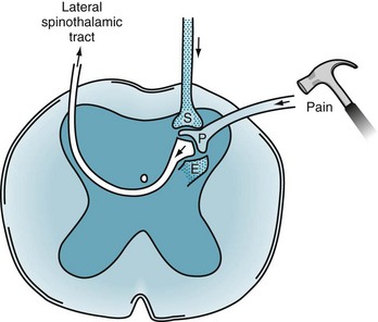
After the synapse, pain sensation ascends predominantly within the lateral spinothalamic tract to the brain (see Figs 2-6 and 2-15). This crucial tract crosses from the substantia gelatinosa to the spinal cord’s other side and ascends, contralateral to the injury, to terminate in specific thalamic segments. Additional synapses relay the stimuli to the somatosensory cerebral cortex, enabling the individual to locate the pain.

In the spinohypothalamic tract, another ascending pain pathway, ipsilateral and contralateral fibers travel up the spinal cord and terminate directly in the hypothalamus. This pathway may explain pain-induced disturbances in temperature regulation, sleep, and other autonomic functions. The spinal cord also transmits pain in other, less well-defined ipsilateral and contralateral tracts.

In addition to relaying pain to the thalamus and hypothalamus, these tracts convey pain to the limbic system, reticular activating system, and other brainstem regions. These connections partially explain why individuals awaken when given a painful stimulus during sleep. It also accounts for chronic pain patients’ sleeplessness, loss of appetite, and a tendency to develop anxiety and mood disturbances. On the other hand, loss of this connection – from trauma, anoxia, disease, or other insult – explains why patients who are unconscious cannot experience pain or suffer.

Analgesic Pathways

Many analgesic pathways interfere with pain transmission within the brain or spinal cord. Several pathways that originate in the frontal lobe and hypothalamus terminate in the gray matter surrounding the third ventricle and aqueduct of Sylvius (periaqueductal gray matter). They contain large amounts of endogenous opioids, which are powerful analgesics (Box 14-1). Implanted electrodes that stimulate the periaqueductal gray matter area may provoke the release of endogenous opioids and thereby induce profound analgesia.

Similarly, short neurons located entirely within the spinal cord, interneurons, inhibit incoming PNS stimuli. These neurons release endogenous opioids and other neurotransmitters that reduce pain transmission.

Analgesic pathways also originate in the brainstem and descend in the spinal cord’s dorsolateral funiculus. They provide “descending analgesia” relief of pain by inhibiting both spinal cord synapses and their ascending pathways. Unlike most other analgesic pathways, they release serotonin.

Endogenous Opioids

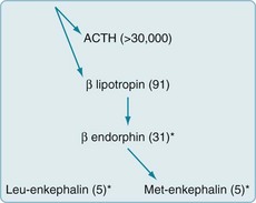
Often called endorphins (endogenous morphine-like substances), endogenous opioids – endorphins, enkephalins, and dynorphins – are powerful analgesic, amino-acid chains (polypeptides) synthesized in the CNS (Fig. 14-2). Endogenous opioids bind to receptors in the limbic system, periaqueductal gray matter, dorsal horn of the spinal cord, and other CNS sites. Neurologists commonly say that a “runner’s high” and the initial painlessness reported by wounded soldiers serve as examples of endorphins’ analgesic effects.
Synthetic (exogenous) opioids, particularly morphine and other medicines, are virtually identical to endogenous opioids. They bind to the same CNS receptors and produce the same effects – analgesia, mood elevation (euphoria), sedation, and respiratory depression. Naloxone (Narcan), an antagonist that competitively binds to the opiate receptor, reverses the effects of endogenous, as well as exogenous, opioids. Indeed, naloxone’s opioid antagonist effect is so characteristic that naloxone-reversibility serves as a criterion for ascertaining that opioid pathways mediate an analgesic’s effect.

Treatments

Physicians prescribe numerous medications and administer them through various routes. Some alleviate pain by reducing tissue damage, interrupting pain transmission through peripheral or central pathways, or blunting its impact on cerebral structures. The addition of psychologic treatment and physical therapy may further reduce pain and, as part of a complete care plan, decrease suffering, restore activities of daily living, and return control to patients. Because chronic pain remains notoriously refractory to conventional treatment, neurologists aim for pain management, not pain cure.

Nonopioid Analgesics

As previously mentioned, aspirin, other salicylates, NSAIDs, steroids, and acetaminophen – nonopioid analgesics – inhibit prostaglandin synthesis at the injury (Box 14-2). Through this mechanism, these medicines relieve acute and chronic pain of mild to moderate severity.

Nonopioid analgesics generally provide steady analgesia for weeks to months and avoid several potential problems. In particular, after completing a course of treatment, patients do not experience withdrawal symptoms. Also, except for high-dose steroids potentially causing steroid psychosis (see Chapter 15), these analgesics do not induce mood, cognitive, or thought disorders.

On the other hand, large doses of NSAIDs and aspirin cause gastric irritation or hemorrhage, and prolonged use increases the risk of cardiovascular disease. In addition, although nonopioid analgesics provide dose-dependent pain relief, they do so only up to a point. Once these medicines provide their maximum pain relief, greater doses do not increase their benefit – the “ceiling effect.”

Nonopioid analgesics are more effective if patients take them on a prophylactic basis. For example, taking nonopioid analgesics prior to dental procedures or menses will avert much of the pain. They are also more effective if taken in a generous initial “loading” dose.

Although nonopioid analgesics alone offer minimal benefit in cases of neuropathic pain, they act synergistically when taken in combination with opioids. In other words, peripherally acting nonopioid analgesics enhance centrally acting opioid analgesics. For example, adding NSAIDs to morphine helps alleviate the pain of metastases to bone. Because nonopioid analgesics allow a smaller dose of opioids to be effective, they have an “opioid-sparing effect.”

With routine use, acetaminophen by itself or as an enhancement to opioids provides effective, reasonable safe analgesia. Stern warnings about acetaminophen compounds, such as oxycodone-acetaminophen (Percocet) and hydrocodone-acetaminophen (Vicodin), as well as acetaminophen itself, causing permanent liver damage are justifiable, but usually when looking at the aftermath of an overdose or their use by alcoholic individuals.

Opioids

When chronic pain results from cancer, neurologists categorize it as “cancer” or “malignant” pain, but when it results from other conditions, “noncancer” pain. Opioids are unquestionably indicated for cancer pain (see Box 14-2). In addition, a number of studies suggest that they are indicated for chronic noncancer pain syndromes.

Opioids have no ceiling effect. Greater doses or more potent preparations increase their analgesic effect. Adding NSAIDs or other nonopioid analgesics enhances opioids’ effect without risking their side effects. On the other hand, when treating patients with noncancer pain who do not find relief with opioid treatment, physicians should slowly discontinue opioids rather than increasing the dosage.

Chronic pain patients may obtain opioid treatment from transdermal patches, intranasal sprays, rectal suppositories, and epidural injections. A particularly innovative technique, patient-controlled analgesia (PCA), allows patients to regulate continual or intermittent intravenous opioid infusions. Through controls in the system, patients regulate the depth of analgesia without causing respiratory depression. Even 6-year-old children can safely and effectively administer PCA.

Compared to older opioids, newer ones provide more rapid onset and longer duration of action. Some are long-acting because they are embedded in a matrix that slowly releases its medication.

Administering opioids by PCA, patches, or long-acting oral preparations on a regular prophylactic or time-dependent basis, such as every 2–4 hours, is more effective than administering them only at the onset of pain. By administering opioids only after pain has developed makes it more difficult to control, creates a pattern of “hills and valleys” (first undertreatment and then overtreatment), prevents a restful sleep, and increases side effects. Moreover, because patients, fearful about pain recurrence, develop anxiety and preoccupation with obtaining their medicines, they may seem to behave as addicts (see later).

Physicians who prescribe narcotics should select long-acting preparations, such as methadone or a transdermal opioid. Also, because various opioids affect different regions of the mu (µ) and related receptors, physicians should vary the opioid for patients with intractable pain. Transdermal and extended-release oral opioid formulations carry a disproportionately great risk of morbidity and mortality.

Physicians should also avoid changing from oral to parenteral forms of opioid at a given dose because the substitution will likely lead to an overdose. In the reverse situation, changing the same dose from an intramuscular or intravenous injection to pills is likely to produce undertreatment, which would cause withdrawal symptoms and recurrence of pain.

In an attempt to control indiscriminate prescription of opioids, the Food and Drug Administration will soon launch Risk Evaluation and Mitigation Strategies (REMS) for physicians who prescribe opioids. REMS will hopefully reduce inappropriate prescriptions, abuse, overdose, and other untoward effects.

“Addiction”

In a situation akin to drug addicts, within weeks of beginning opioid treatment, patients require increasingly greater quantities to produce the same level of anesthesia (tolerance) as opioids desensitize receptors. Similarly, abruptly stopping opioid treatment produces unpleasant symptoms (withdrawal). However, because the two situations differ in several important respects, physicians certainly do not label a terminal cancer patient who requires opioids a drug addict.

Although tolerance and withdrawal characterize physical dependence, physicians in pain management services, as ones in psychiatry, define addiction primarily in behavioral terms, such as potentially harmful drug-seeking activity and overwhelming involvement with use of a drug. They consider tolerance and dependence, which often occur together but can occur independently, as physiologic responses, an expectable aspect of medical treatment, and not peculiar to opioids. They also note that inadequate treatment and development of tolerance drive pain patients to be overly concerned about their opioid schedule and to seek larger doses. Physicians unfamiliar with pain management may interpret this behavior as “drug-seeking” and an indication of addiction, but many pain management physicians consider it iatrogenic pseudoaddiction. On the other hand, some pain management physicians remain skeptical and consider pseudoaddiction to be true addiction.

Moreover, pain management physicians quip that other physicians’ fear of creating addiction has led to the most common side effect of opioid treatment, undertreatment. In fact, addiction rarely develops in previously opioid-naive individuals who develop an acute painful illness that requires opioids for several weeks. Pain management physicians have been advocating continual opioid treatment of moderate to severe noncancer as well as cancer pain.

Other Opioid Side Effects

For the psychiatric consultant, a dilemma is the similarity of opioid-induced mental status changes to those induced by head trauma, metabolic aberrations, and cerebral metastases. Because opioids may cause delirium and depress the level of consciousness, which would obscure the clinical picture, neurologists and neurosurgeons avoid treating head trauma patients with opioids.

Another opioid side effect, hypoventilation – slow and shallow breathing – represents its primary life-threatening one. However, in practical terms, administering opioids to patients with pulmonary disease, rather than an overdose, is the usual cause of hypoventilation. Also, patients develop tolerance to hypoventilation along with their other side effects.

Although depression of sensorium and hypoventilation loom large to physicians as major iatrogenic problems, constipation remains the most troublesome side effect for patients. Rather than trying to reverse this inevitable complication, physicians can prevent it by ordering a combination of laxatives, such as senna (Senokot), and stool softeners, such as docusate sodium (Colace). They can also prescribe an oral, selective mu-opioid receptor antagonist that does not cross the blood–brain barrier, such as alvimopan (Entereg).

To prevent or alleviate opioid-related nausea, physicians should prescribe antiemetics, but they should cautiously prescribe antiemetics containing phenothiazine or other dopamine-blocking agent because they can cause dystonic reactions or parkinsonism (see Chapter 18). In another caveat, pharmacologic marijuana, such as dronabinol (Marinol) and nabilone (Cesamet), generally reduces nausea, pain, and anxiety; however, robust formal studies have not established that they are the most effective treatment when cancer or chemotherapy has been responsible for these symptoms. Moreover, they may cause transient mood and thought disorders.

The use of specific opioids is fraught with difficulties. For example, phenytoin and carbamazepine – whether prescribed for their antiepileptic activity, mood modulation, or pain control – accelerate methadone metabolism. Therefore, giving either of these AEDs to patients on methadone maintenance may precipitate withdrawal symptoms. To avoid that problem, physicians should increase the methadone dose when they add those medications.

Heroin, a problematic opioid, is no more effective in relieving pain or improving mood than morphine, but its potential for abuse is much greater. Regardless of several medical and nonmedical groups’ assertions, heroin has no legitimate use that cannot be better fulfilled by the current array of medicines.

As with other medicines, physicians should discontinue opioids when unnecessary. Rather than abruptly stopping opioids, which would probably lead to withdrawal, physicians might use an exit strategy of tapering opioids by reducing their dose by 50% every 3 days. Alternatively, physicians might briefly replace a short-acting opioid, such as morphine, with a long-acting one, such as methadone, and then prescribe a nonopioid analgesic. If withdrawal still complicates the process, benzodiazepines may alleviate the physical or mental discomfort, and clonidine (Catapres) may blunt autonomic nervous system hyperactivity (see Chapter 21).

Chronic Opioid Therapy Debate

Over the past decade, neurologists, pain management specialists, and other physicians began to prescribe long-term opioid treatment for many noncancer conditions, including osteoarthritis, headache, postoperative low back pain, and fibromyalgia. These physicians offer several arguments that have gained increased acceptance. They claim that, when used to control otherwise intractable pain, opioids are not addictive; opioids are effective, safe, and allow patients to work, use machinery, and drive; and, more than other medicines, opioids increase function, reduce suffering, and restore sleep.

Physicians prescribing chronic opioid therapy expect patients to agree, often with a written contract, to use only the opioid in preset amounts and only from a single physician. Patients must also give permission for random urine testing for illicit substances, particularly drugs of abuse. Physicians monitor patients’ social and occupational functioning as well as their pain and urine tests.

Physicians who have opposed expanding opioid prescription offer several counterarguments. Opioids reduce pain less than 2–3 points on a 0–10 scale. Chronic opioid therapy may be counterproductive in several conditions. It portends worse long-term outcomes for headache and low back pain, and tends to convert episodic migraines or tension-type headache to persistent daily headache (see Chapter 9). Patients demand opioids when less potent or alternative measures, even nonpharmacologic ones, would suffice. In addition, patients may subvert medical treatment. They may seek opioids for their euphoric effect rather than for pain relief. Falsely reporting the persistence and severity of pain to obtain opioids, patients prolong their disability. Some patients, claiming intractable pain, demand excessive quantities, but sell or pass along their opioids. To avoid this “diversion” of opioids, some physicians go so far as to ask the patient who has been prescribed fentanyl patches to return the used ones, which should have the patient’s hair stuck to the patch’s adhesive. Some patients use opioids to tide them over an addiction to illicit drugs. For many patients, obtaining opioids takes over their day-to-day concerns.

Some neurologists feel that chronic opioid therapy is rife with misuse. Examples include using other alcohol and illicit substances in addition to the opioids, selling or trading the opioids, and surreptitiously obtaining additional quantities of opioids. Risk factors for misuse are family as well as a personal history of substance abuse; age 16–45 years; presence of a “psychiatric condition,” which includes schizophrenic disorders, affective psychosis, neurotic disorders, and personality disorders; and, in women, preadolescent sexual abuse. Rates of misuse of opioids among chronic opioid therapy patients range from 3% to 30%.

Long-term opioid treatment, especially at high dosage or following a dose increase, generally produces sedation, impairs cognition, and interferes with psychosocial function. In addition to leading to these neuropsychologic effects, it leads to the usual opioid side effects, particularly chronic constipation and hypogonadism.

Finally, physicians prescribing opioids are subject to administrative oversight that may be stringent and punitive. Both state and federal investigators pursue possible irregularities.

Adjuvants

Other Treatments Directed at the Peripheral or Central Pathways

When the skin is affected by postherpetic neuralgia (see later) or other painful conditions, applying patches of a long-acting anesthetic agent, such as lidocaine gel (Lidoderm), directly over the lesion interrupts the transmission of pain stimuli. Alternatively, for a region of occipital scalp, chronic chest, or abdominal pain, physicians may perform a nerve block by injecting anesthetic agents into one of the cervical, thoracic, and lumbar nerve roots, which are readily accessible as they emerge from the spine.

Long-acting local preparations provide pain relief for days or, with an alcohol, months. However, nerve blocks are impractical for chronically painful limbs because the blocks may cause paresis, as well as analgesia. Nor are they useful for facial pain within the first division of the trigeminal nerve (V1) because analgesia involving the eye leads to corneal ulceration.

Sometimes a sympathetic plexus or ganglion block reduces pain. For patients with pancreatic carcinoma, physicians can deaden the celiac plexus with alcohol injections. Similarly, for patients with the shoulder–hand syndrome, physicians can inactivate the stellate ganglion (the sympathetic ganglion adjacent to the upper cervical vertebrae) with alcohol injections. Nevertheless, their benefit in complex regional pain syndrome remains controversial.

To interrupt pain transmission in the spinal cord, physicians have introduced several different treatments. Capsaicin cream, applied to a painful area, is absorbed through the skin and drawn up along sensory nerves. When it reaches the spinal cord synapse, capsaicin depletes substance P, the crucial neurotransmitter, and thereby impairs pain transmission (see Fig. 14-1). This treatment, which complements systemic medicines, helps alleviate pain from arthritis, diabetic neuropathy, and postherpetic neuralgia.

Anesthetics administered intrathecally, the basis of spinal anesthesia, completely block transmission of all sensory and motor nerve impulses. Although suitable for surgical procedures, spinal anesthesia is not selective enough for the treatment of chronic pain. Preliminary studies suggest that certain intrathecally administered nonopioid voltage-gated calcium channel blockers, such as ziconotide, create anesthesia without producing paresis or incontinence. Another approach to interrupting spinal cord pain transmission, dorsal column stimulation, applies a low-intensity current to the spinal cord (see later).

Stimulation-Induced Analgesia

The idea that stimulation of a neurologic pathway inhibits a complementary one has given rise to stimulation-induced analgesia. The gate control theory, which embodies this idea, supposedly found practical application in transcutaneous electrical nerve stimulation (TENS). TENS devices, which stimulate the skin proximal to the painful area, generate low-intensity impulses that theoretically dampen adjacent pain-transmitting ones. Despite their widespread use, with many pain patients reporting substantial relief without side effects, studies have shown no benefit for their most popular application, chronic low back pain, but some relief for painful diabetic neuropathy.

A similar but invasive technique, spinal (dorsal column) cord stimulation, involves neurosurgeons inserting electrodes into the epidural space overlying the spinal cord. A stimulator, which the patient controls, generates an electric current that presumably interferes with nerve transmission. According to preliminary reports, the device alleviates pain and improves the quality of life for patients with complex regional pain syndrome or chronic low back pain.

Along the same line, some studies found that acupuncture provides analgesia in people with mild to moderate pain. Placing the needles in dermatomes (see Fig. 16-2) creates more analgesia than placing them in the traditional regions (meridians). When acupuncture includes electrostimulation, the procedure doubles its effectiveness. Because traditional acupuncture induces a rise in brainstem, limbic system, and cerebrospinal fluid endorphins and its benefit is naloxone-reversible, acupuncture may work in part through the endogenous opioid system (Box 14-3).

In another CNS stimulation technique, neurosurgeons implant electrodes into the periventricular and periaqueductal gray matter and adjacent brainstem regions. As noted earlier, stimulation of these sites, which releases stored endogenous opioids, may induce profound analgesia. Despite the strong rationale, the procedure remains investigational.

For patients with intractable pain confined to a single limb, neurosurgeons have experimented with severing the lateral spinothalamic tract in the spinal cord contralateral to the pain. This procedure, a cordotomy, based on the neuroanatomy of the spinothalamic tract, provided profound and almost immediate analgesia. However, because the improvement lasted for only a few months, physicians abandoned the procedure. Neurologists attributed the brevity of its effect to plasticity of central pain pathways. Bilateral cordotomies for extensive pain were also unacceptable because they were complicated by respiratory drive impairment (Ondine’s curse) and urinary incontinence.

Epidural Analgesia

Anesthesiologists administer epidural analgesia, an epidural, for vaginal birth, cesarean section, or other pelvic surgical procedures. Epidurals anesthetize nerve roots T10 through L1, which innervate the uterus and cervix, and S2–4, which, through the pudendal nerves, innervate the vaginal canal and perineum (see Fig. 16-1). They create analgesia throughout the pelvis, but do not cause respiratory depression in the woman or affect the fetus.

The procedure typically consists of the anesthesiologist introducing a catheter into the lumbar epidural space and injecting a local anesthetic, such as lidocaine, along with an opioid anesthetic, such as morphine (Fig. 14-3). The anesthetics permeate the lumbosacral epidural space, but, to a limited extent, they also diffuse into the underlying subarachnoid space. Depending on the duration of the procedure, required depth of analgesia, and other factors, the anesthesiologist continually administers anesthetics through the catheter. When the anesthetic bathes only the lowermost nerves, as for delivery and other obstetric procedures, patients retain strength in their legs and remain able to walk.
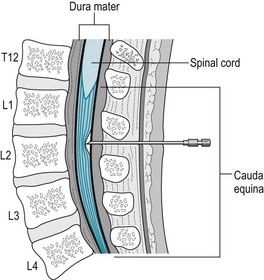
A related procedure, spinal anesthesia, a spinal, may supplement an epidural. In this procedure, the anesthesiologist injects anesthetic directly into the lumbar subarachnoid space. This technique, which is similar to a lumbar puncture, provides rapid-onset anesthesia. It also causes paraparesis. However, the anesthetic may diffuse upward in the subarachnoid space to compromise the spinal cord. If the anesthetic rises to the cervical region, it may cause quadriplegia and respiratory arrest.

Cancer Pain

Cancer can cause neuropathic and nociceptive pain that, if improperly treated, may be excruciating and unrelenting. Such pain may originate in a variety of sources, particularly from metastases in bones, which are richly innervated, and nerves. The medical and surgical treatments themselves may also cause pain. Moreover, disease-induced and iatrogenic pain superimposed on the underlying illness routinely engenders depression and anxiety.

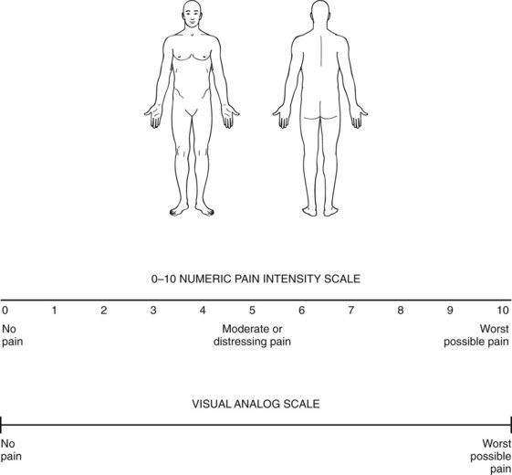
Physicians should generally accept patients’ accounts of their pain without reservation and monitor their pain as regularly as they check the temperature and pulse (Fig. 14-4). In fact, pain has come to be regarded as the “fifth vital sign.” Patients should have access to nonpharmacologic treatments as well as the full arsenal of medicines. Physicians should prescribe medicines from the three categories – nonopioid, opioid, and adjuvant – early, if not pre-emptively, and then frequently and generously. As opioid tolerance develops, physicians should readily increase the doses. In addition, they should suppress “breakthrough pain” with supplements to regularly scheduled opioids.
Noncancer Pain Syndromes

Although agonizing pain is generally associated with malignancies, several noncancer syndromes cause comparable pain, suffering, and disability. In addition, insomnia, drug-seeking behavior, depression, and other psychiatric comorbidities complicate these syndromes.

Neurologists often successfully apply guidelines for the management of cancer-related pain to noncancer pain syndromes. Nevertheless, some differences exist. Patients and physicians should acknowledge that, while most of these conditions are chronic and incurable, they are not fatal. The goals in these treatment strategies – different from the single-minded one of complete pain relief – should be to reduce suffering, restore ability to work, and allow a return to social roles. The goals should be clear, acceptable to the patient and caregivers, and attainable within several months.

Physicians should strive to control, if not alleviate, pain. The treatment plan, which usually requires patient and family cooperation, should aim to minimize suffering, medication side effects, and preoccupation with pain and obtaining medications. Medications should maintain patients’ mental clarity and function.

The book has already reviewed several common noncancer pain syndromes, including diabetic neuropathy (see Chapter 5) and trigeminal neuralgia (see Chapter 9). This chapter continues by discussing six others. Their pharmacologic management often begins with analgesics, which often includes chronic opioids. TCAs, SNRIs, gabapentin, and pregabalin are more effective in the treatment of neuropathic than nociceptive pain. Patients may still require opioids. Physical and occupational therapy, psychotherapy, hypnosis, behavioral therapies, and social service intervention may further reduce suffering, improve mobility, and increase function.

Postmastectomy Axillary Pain

During a mastectomy, as surgeons explore the axilla and remove lymph nodes, they occasionally damage or sever the cutaneous branch of the first thoracic nerve root, the intercostobrachial nerve. Several weeks after the surgery, some women develop searing axillary pain that extends to the inner aspect of the upper arm, well beyond the incision. (In contrast, common incision pain is only mildly to moderately intense, has an itching quality, and is confined to the scar.) Postmastectomy pain, like many painful conditions, worsens at night. Because shoulder movement provokes postmastectomy pain, pain-induced immobility may lead to a “frozen shoulder.”

Approximately 50% of women undergoing mastectomy will suffer postoperative pain and 10% will describe it as “moderate to severe.” Neurologists attribute postmastectomy pain to intercostal-brachial neuralgia, phantom breast pain, pain related to implants and other surgical reconstruction, and peripheral neuropathy. Some unfortunate women may develop similar pain if they have a cancer recurrence with metastatic invasion of the brachial plexus. Risk factors for postmastectomy pain include preoperative chronic pain, repeated surgery, outstanding litigation, surgery that involved axillary node dissection, breast-conserving procedures, and insertion of implants.

Opioid or nonopioid analgesics and adjuvants usually help women with routine postmastectomy pain. Local treatments, such as lidocaine gel or nerve blocks, provide topical anesthesia that complements systemic treatment. In addition, physical measures, such as massage, shoulder exercises, and vapor coolants, e.g., ethyl chloride spray, may reduce the pain.

Even though postmastectomy pain carries little prognostic significance, it intensifies the surgery’s psychologic impact. Moreover, it tempts psychologically oriented physicians and nonphysicians to suggest that postmastectomy pain stems from emotional factors.

Postherpetic Neuralgia

An acute herpes zoster infection, from reactivated varicella virus that has lain dormant in dorsal (sensory) nerve root ganglia, changes one or two adjacent dermatomes into a field of vesicular eruptions, shingles. The infection causes an unrelenting, moderately severe burning sensation that may precede eruption of the vesicles by several days. This pain typically lasts several weeks. Although the infection may strike any dermatome, it most commonly affects the thoracic dermatomes and the first branch of the trigeminal nerve, which includes the cornea. It causes pain not only during several weeks of the acute infection but then, in many cases, a recurrence involving chronic severe pain, i.e., the pain often has a bimodal course.

Herpes zoster virus commonly infects individuals older than 65, even those in good health, and patients with immunosuppressive illnesses, such as lymphoproliferative disorders and acquired immunodeficiency syndrome (AIDS). A vaccine against herpes zoster may prevent, reduce the incidence, or at least decrease the severity of the infection and all of its painful sequelae. Because one-third of all people may experience a herpes zoster infection during their lifetime, this vaccine has the capacity to produce a public health improvement.

The pain of acute herpes zoster infection clearly merits a prescription of an opioid. Antiviral agents, such as acyclovir (Zovirax) and famciclovir (Famvir), may speed healing of vesicles, shorten duration of the pain, and prevent spread of infection to the eye.

For the 3–6 months after the initial infection has resolved, patients are at risk of developing an even more painful secondary phase, postherpetic neuralgia. In an age-related risk pattern, postherpetic neuralgia develops in few individuals younger than 50 years, but in 50% of those older than 60 years and in 75% of those older than 70 years.

The neuralgia has a continual dull quality of excruciating severity with superimposed lancinating paresthesias. The agony it causes leads to anorexia, insomnia, and mood changes. Although postherpetic neuralgia eventually resolves spontaneously, it may torture patients for years and leave them with a band of anesthetic (numb) scarred skin and, if a major motor nerve is involved, muscle weakness and atrophy. Physicians should prescribe adjuvants, particularly gabapentin and pregabalin, as well as generous doses of long-acting opioids. If the affected area does not include the eye, patients might also benefit from long-acting topical anesthetics, such as lidocaine gel. As a general rule, the earlier the diagnosis and treatment of postherpetic neuralgia, the more likely treatment is to be effective.

Complex Regional Pain Syndrome

Reflex sympathetic dystrophy (RSD), causalgia, and sympathetically maintained pain fall under the rubric of complex regional pain syndrome. By definition, type 1 complex regional pain syndrome stems from no definable nerve injury, but type 2 has an underlying definable nerve injury. However, terms suggesting that the sympathetic nervous system is at fault are misleading because studies have demonstrated no consistent abnormalities in it.

The severity, chronicity, and disability of patients’ pain are typically much greater than expected for the injury. In addition, the pain often extends far beyond the injury to include the entire limb, although predominantly in its distal portions, and in many cases into adjacent limbs. For example, after several months of left arm pain, a patient may describe similar symptoms in the left leg and then in the right leg.

Patients typically describe the pain as relentless burning or lancinating sensations superimposed on irritating numbness. Sometimes their descriptions of the pain seem extravagant. Neurologists find allodynia, hyperalgesia, and hyperpathia in complex regional pain syndrome patients more often than in any of the other noncancer pain syndromes. Because even touching or moving an affected limb increases the pain, patients tend to be preoccupied with the affected limb and go to great lengths to guard it. For example, they assiduously shield an afflicted hand by wearing a sling and glove, or they protect a foot by using crutches. The combination of the injury, sensitivity, and protective maneuvers incapacitates the affected limb.

Fingernails remain uncut and grow thick, long, and brittle. Although the skin may sweat excessively (hyperhidrosis), it is usually smooth, shiny, scaly, and dry. Similarly, the skin of affected regions usually turns pale, sometimes changing to a dusky color with a superimposed livedo reticularis. Severe cases include peripheral edema, bone reabsorption, and muscle wasting (dystrophy), which gave rise to the term RSD. Tremors, dystonia, and other involuntary movement disorders complicate some cases.

Complex regional pain syndrome follows minor as well as major injuries of a limb. The injuries may not necessarily have involved overt nerve damage, as with a penetrating wound. Compressive or traction injuries may have preceded its development. Common causes are knife or gunshot wounds, fractures, myocardial infarctions, and peripheral vascular disease.

In addition to prescribing the standard treatments for neuropathic pain, physicians order intensive physical therapy to mobilize the affected limb. They also prescribe blockade of regional sympathetic ganglia in selected cases. For example, patients with causalgia of the hand may benefit from blocking the stellate ganglion with an anesthetic agent. Intravenous infusions of bretylium or guanethidine, which block the region’s α- and β-adrenergic sympathetic ganglion receptors, occasionally produce dramatic pain relief. Response to such blocks, even though brief, has diagnostic as well as therapeutic value.

Physicians also attempt to block the pain pathways in the spinal cord by implanting spinal stimulators, but the procedures are invasive and dangerous, and remain experimental. Similarly, ziconotide, the calcium channel blocker, holds promise as a medication that will interrupt the spinal cord pain pathways.

Phantom-Limb Pain

Phantom-limb pain occurs in areas that had been a limb before it was amputated. For example, a man with this disorder, who lost his left leg at the thigh when it was severed in an automobile accident, still felt pain in his left ankle for many months. In addition, nonpainful sensations often accompany painful ones. For example, patients with an amputation will often feel that their limb remains an integral part of their body and sense it making purposeful movements. The phenomena are not restricted to limb injuries but may follow amputation of a breast, ear, or other body part.

Neurologists often classify the pain from phantom limb, brachial plexus avulsion, and thalamic infarction (see later) as deafferentation pain because sensory deprivation underlies all of these conditions. Whatever their origin, abnormal sensations or misperceptions reach the level of somatic hallucinations. They resemble the visual hallucinations that stem from visual deprivation, i.e., blindness.

Pain at the site of a surgical incision, stump pain, differs from phantom-limb pain because it is confined to the injury and attributable to nerve scars (neuromas). Nevertheless, phantom and stump pain may occur together.

Even in its most typical settings, phantom-limb pain varies in quality, severity, and accompanying psychologic symptoms. It usually begins soon after a traumatic amputation and has a self-limited duration of several weeks. The pain is even more likely to develop and persist if the affected body part were chronically painful before the amputation (as with osteomyelitis). Because most cases result from war wounds, victims are usually young or middle-aged veterans. Many patients also have sustained other injuries, including facial disfigurement, loss of several limbs, and castration.

In addition to prescribing the usual treatments for noncancer pain syndromes, some physicians have claimed to help patients using hypnosis that induces the imagery that the phantom limb is shrinking to the point of disappearing. Another nonpharmacologic approach is “mirror therapy,” which induces changes in the primary sensorimotor and supplementary motor cortex.

Low Back Pain

As opposed to acute low back pain from a herniated intervertebral disk (see Chapter 5), chronic low back pain seems to stem from ill-defined or nonspecific injury of the lumbar vertebrae, intervertebral disks, or the supporting ligaments. Affecting over 2 million individuals, chronic low back pain is one of the most prevalent disabling conditions in the United States.

As much as in any neurologic condition, nonmedical factors contribute to persistent pain and prolonged disability. In fact, disability bears little relationship to structural abnormalities visible on magnetic resonance imaging (MRI). Work-related injuries, job dissatisfaction, psychological distress, outstanding litigation, and other painful conditions are risk factors for a poor prognosis. Although depression, distress, somatization, and substance abuse also play a major role, their effects are often hidden. Psychologic screening tests, including the Minnesota Multiphasic Personality Inventory-2 (MMPI-2), cannot reliably predict which chronic low back pain patients will have a satisfactory surgical outcome.

Most patients with chronic low back pain should be advised to accept it as a chronic condition that medical care might ameliorate but not cure. Physicians might shift their goal from abolishing pain to improving function. For example, goals, which should be kept modest, might include completing a 6-hour work day or walking 1 mile (1.6 km).

Although the usual treatments for noncancer pain help, severe cases defy treatment. For example, despite even obvious MRI abnormalities, surgery on herniated disks, spinal stenosis, and other orthopedic conditions fails in the majority of cases to eliminate pain and enable patients to return to work. In fact, surgery may even worsen the pain and disability. Also, many popular techniques – acupuncture, back-strengthening exercises, massage, spinal manipulation, and sleeping on a firm mattress – produce mild, short-lived, or no improvement.

Neurologists often refer to patients who remain incapacitated despite surgery and other treatment as having the “failed back surgery syndrome” (FBSS). Studies of these patients have postulated multiple causes of FBSS, such as neural fibrosis and recurrent or retained herniated disks, but have shown no explanation in a significant proportion of cases.

Finally, both physicians and patients should acknowledge, when relevant, that litigation often promises large amounts of money for permanent pain, suffering, and disability. This incentive discourages improvement, effective treatment, a return to work, and even accurate reporting. Some neurologists recommend concluding litigation before expecting successful treatment.

References

Abramowicz M. Medical marijuana. Med Lett. 2010;52:5–6.

Backonja MM, Krause SJ. Neuropathic pain questionnaire – short form. Clin J Pain. 2003;19:315–316.

Ballantyne JC, Mao J. Opioid therapy for chronic pain. N Engl J Med. 2003;349:1943–1953.

Bartleson JD. Evidence for and against the use of opioid analgesics for chronic nonmalignant low back pain: A review. Pain Med. 2002;3:260–271.

Benarroch EE. Sodium channels and pain. Neurology. 2007;68:233–236.

Berman BM, Langevin HH, Witt CM, et al. Acupuncture for chronic low back pain. N Engl J Med. 2010;363:454–461.

Carragee EJ. Persistent low back pain. N Engl J Med. 2005;352:1891–1898.

Carragee EJ, Alamin TF, Miller JL, et al. Discographic, MRI, and psychosocial determinants of low back pain disability and remission: A prospective study in subjects with benign persistent back pain. Spine J. 2005;5:24–35.

Carroll LJ, Cassidy JD, Cote P. Depression as a risk factor for onset of an episode of troublesome neck and low back pain. Pain. 2004;107:134–139.

Cherkin DC, Sherman KJ, Deyo R, et al. A review of the evidence for the effectiveness, safety and cost of acupuncture, massage therapy, and spinal manipulation for low back pain. Ann Intern Med. 2003;138:898–906.

Diers M, Christmann C, Koeppe C, et al. Mirrored, imagined and executed movements differentially activate sensorimotor cortex in amputees with and without phantom limb pain. Pain. 2010;149:296–304.

Dubinsky RM, Miyasaki J. Practice parameter: Treatment of postherpetic neuralgia. Neurology. 2004;63:959–965.

Dubinsky RM, Kabbani H, El-Chami Z, et al. Assessment: Efficacy of transcutaneous electric nerve stimulation in the treatment of pain in neurologic disorders (an evidence-based review). Neurology. 2010;74:173–176.

Dworkin RH, O’Connor AB, Audette J, et al. Recommendations for the pharmacological management of neuropathic pain: an overview and literature update. Mayo Clin Proc. 2010;85(3 Suppl):S3–S14.

Foley KM. Opioids and chronic neuropathic pain. N Engl J Med. 2003;348:1279–1281.

Hawkins JL. Epidural analgesia for labor and delivery. N Engl J Med. 2010;362:1503–1510.

Hayden JA, van Tulder MW, Malmivaara AV, et al. Meta-analysis: Exercise therapy for nonspecific low back pain. Ann Intern Med. 2005;142:765–775.

Jovey RD, Ennis J, Gardner-Nix J, et al. Use of opioid analgesics for the treatment of chronic noncancer pain – a consensus statement and guidelines from the Canadian Pain Society, 2002. Pain Res Manag. 2003;Suppl A:3A–28A.

Jung BF, Ahrendt GM, Oaklander S, et al. Neuropathic pain following breast cancer surgery. Pain. 2003;104:1–13.

Nasreddine ZS, Saver JL. Pain after thalamic stroke: Right diencephalic predominance and clinical features in 180 patients. Neurology. 1997;48:1196–1199.

Nicholson B, Passik SD. Management of chronic noncancer pain in the primary care setting. South Med J. 2007;100:1028–1036.

Pincus T, Burton AK, Vogel S, et al. A systemic review of psychological factors as predictors of chronicity/disability in prospective cohorts of low back pain. Spine. 2002;27:109–120.

Rowbotham MC, Twilling L, Davies PS, et al. Oral opioid therapy for chronic peripheral and central neuropathic pain. N Engl J Med. 2003;348:1223–1232.

Sampathkumar P, Drage L, Martin D. Herpes zoster (shingles) and postherpetic neuralgia. Mayo Clin Proc. 2009;84:274–280.

Slipman CW, Shin CH, Patel RK, et al. Etiologies of failed back surgery syndrome. Pain Med. 2002;3:200–214.

Webster LR, Webster RM. Predicting aberrant behaviors in opioid-treated patients. Pain Med. 2005;6:432–442.

Williams LS, Jones WJ, Shen J, et al. Outcomes of newly referred neurology outpatients with depression and pain. Neurology. 2004;63:674–677.

Answer:

b. Pseudoaddiction refers to a patient’s concern to the point of obsession with securing a pain-relieving medication, hoarding it, and other addiction-like behavior. Provided that the patient receives adequate medication at the appropriate dose and time, patients’ behavior should be normal. On the other hand, the patient may have developed addiction once he began opioid treatment or even before. Alternatively, he may be self-medicating for depression, anxiety, personality disorder, or other psychiatric disorder. Several medications, particularly phenytoin, may interfere with the opioids. Finally, physicians dispensing opioids must guard against their patients engaging in “criminal diversion,” i.e., selling their narcotic medications.