Neuroblastoma

Published on 06/06/2015 by admin

Filed under Pediatrics

Last modified 22/04/2025

Print this page

rate 1 star rate 2 star rate 3 star rate 4 star rate 5 star
Your rating: none, Average: 5 (1 votes)

This article have been viewed 11087 times

58 Neuroblastoma

Neuroblastoma is the most common extracranial solid tumor of childhood and accounts for 8% to 10% of all childhood cancers. The median age at diagnosis is 17 months, and the vast majority of neuroblastoma is diagnosed before age 10 years. The prognosis varies significantly and depends on age, tumor biology, and extent of disease at diagnosis. Whereas some disseminated tumors in infants may spontaneously regress, metastatic tumors diagnosed in slightly older children carry a long-term survival rate of around 40%. Although many children with neuroblastoma will need aggressive surgical resection and chemotherapy, some children can be safely observed over time. It is this dramatic heterogeneity of phenotypes that has prompted further evaluation of the implications of staging and treatment. Novel therapies are becoming increasingly important in treating refractory disease.

Etiology and Pathogenesis

Neuroblastoma is characterized as a small, round, blue-cell tumor and arises from neural crest cells. The Shimada and Joshi staging system is based on the pathologic components of neuroblastoma. The important histologic components for classification are degree of neuroblast cell differentiation, mitosis-karyorrhexis index (MKI), and amount of stromal content. This combined with age at diagnosis divides patients into two categories: favorable and unfavorable histology.

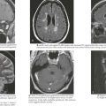
Additionally, a number of genetic aberrations are important in classification and prognosis of neuroblastoma. The DNA content of tumor cells is characterized as near-diploid or hyperdiploid, the latter of which is associated with a more favorable outcome. Examples of other genetic abnormalities associated with prognosis include MYCN amplification, deletion of the short arm of chromosome 1p, and deletion of chromosome 11q. The most important of these, MYCN amplification, is associated with rapid tumor progression and poor prognosis, even in patients with otherwise lower stage disease. Neuroblastoma is most commonly an isolated diagnosis, but it has been associated with other neurocristopathies such as Hirschsprung’s disease, congenital central hypoventilation syndrome (CCHS or Ondine’s curse), and neurofibromatosis type 1. Additionally, a very small number of patients (1%-2%) present as part of a familial neuroblastoma syndrome. In this situation, the most common genetic abnormality is a germline mutation in the anaplastic lymphoma kinase (ALK) gene (Figure 58-1).

Clinical Presentation

Neuroblastoma can arise anywhere along the sympathetic chain, and symptoms at the time of diagnosis depend on the location and extent of disease. Primary tumors most commonly occur in the abdomen (65%) and usually present as a painless abdominal mass. These patients may also present with vomiting, constipation, or symptoms of intestinal obstruction. Other common sites of primary tumors include the neck, chest, and paraspinal region. Tumors arising from the chest may be found incidentally on chest radiography, and cervical tumors may present as a Horner’s syndrome or more rarely with superior vena cava syndrome (Figure 58-2).

Approximately half of patients have metastatic disease at the time of diagnosis. Infants with metastatic disease often present with massive hepatomegaly with or without respiratory compromise. Infants may also present with bluish, subcutaneous nodules, a hallmark of the disease in this population. Other symptoms of metastatic disease in any patient include anorexia, bone pain, irritability, fever, pallor, and hypertension (most often as a result of renal vascular compression). Periorbital ecchymosis and proptosis (from bony tumor infiltrate) are also characteristic of neuroblastoma. Rarely, children are symptomatic from tumor cell catecholamine release, resulting in flushing, sweating, headache, palpitations, and hypertension.

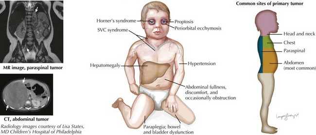
A small percentage of children (5%) present with symptoms of spinal cord compression. This oncologic emergency is associated with paraspinal neuroblastomas, and associated findings include lower extremity weakness, bowel and bladder dysfunction, back pain, and sensory loss. Neurologic function at the time of diagnosis has a strong association with long-term neurologic outcome (see Figure 58-2).

In addition, distinct paraneoplastic syndromes are associated with neuroblastoma. Secretion of vasoactive intestinal peptide by tumor cells results in intractable, watery diarrhea; hypokalemia; and poor growth. These tumors are often associated with favorable histology and good prognosis. The syndrome of opsoclonus–myoclonus is characterized by rapid, involuntary eye movements in all directions; irregular, frequent muscle jerking; and ataxia. These children usually also have a low-stage tumor with favorable biologic features. The presenting neurologic symptoms of opsoclonus–myoclonus and ataxia typically resolve after treatment; however, the majority of children have residual developmental delay.

The previously described clinical symptoms should be carefully evaluated in the physical examination. The abdominal examination should focus on the presence of a mass or hepatomegaly. Head and neck evaluation should include examination for proptosis, periorbital ecchymosis, and Horner’s syndrome (ptosis, miosis, anhidrosis, and ipsilateral facial flushing). One should perform an examination of the cervical, supraclavicular, axillary, and inguinal areas for lymphadenopathy. It is essential to complete a careful neurologic examination because subtle findings can indicate an evolving paraspinal mass. Key neurologic findings include lower extremity weakness, hyperreflexia or diminished weakness, decreased rectal sphincter tone, bowel or bladder incontinence, and paraplegia.

Differential Diagnosis

Because of its varied presentation, the differential diagnosis of a patient with suspected neuroblastoma is broad and should be based on specific signs and symptoms. The clinical presentation can be divided into categories: an abdominal mass or symptoms, a mass in another location, spinal cord compression, and nonspecific neurologic symptoms. Table 58-1 summarizes the differential diagnosis of neuroblastoma using these categories of presenting symptoms.

Table 58-1 Differential Diagnosis of Neuroblastoma

Abdominal Mass
Intraabdominal mass Neuroblastoma Ovarian torsion
Wilms tumor Ovarian tumor
Omphalocele Teratoma
Hernia Lymphoma
Pheochromocytoma Hepatoblastoma
Adrenal hemorrhage Pancreatoblastoma
Hemangioendothelioma  
Intestinal mass Bezoar Intussusception
Volvulus Abscess
Megacolon  
Other intraabdominal pathologies Pancreatic pseudocyst Enlarged uterus
Choledochal cyst Constipation
Hydrops of the gallbladder Cystic kidney disease
Thoracic Mass
Oncologic

Non-oncologic Spinal Cord Compression Oncologic

An abdominal mass is the most common physical examination finding in a patient with neuroblastoma. When evaluating an abdominal mass, it is important to determine whether it is secondary to an enlarged organ (hepatomegaly) or a discrete mass. Even though hepatomegaly may be mistaken for an abdominal mass, an enlarged liver can also be the result of an infection, storage disease, congenital hepatic fibrosis, or malignant infiltration. Splenomegaly may be attributable to leukemia or lymphoma, a hemolytic anemia, portal hypertension, or a storage disease.

There are many causes of spinal cord compression (see Table 58-1) in addition to neuroblastoma. However, in the case of spinal cord compression, the cause may not be immediately known, and steroid treatment can be initiated before a definitive diagnosis is made in select cases. In cases in which neurologic symptoms are rapidly progressing, immediate treatment is warranted to increase the prospect for preservation of neurologic function.

Opsoclonus–myoclonus occurs in 2% to 4% of children with neuroblastoma. It represents a specific syndrome characterized by rapid, irregular eye movements; ataxia; and myoclonus. Approximately 50% of children with this syndrome are found to have a primary neuroblastoma, and diagnostic workup should be undertaken to find a primary tumor in all children with these symptoms. The differential diagnosis is broad for any one of these symptoms; however, the constellation of symptoms significantly narrows the differential diagnosis.

Staging and Management

Staging

The treatment approaches for neuroblastoma depend on a number of clinical and biologic characteristics of the tumor. Tumor risk assessment, as devised by the International neuroblastoma Risk Group, depends on tumor stage, age at diagnosis, and MYCN gene status. These factors along with other biologic variables (mainly genetic aberrations) and tumor histology are used to classify patients into low-, medium-, and high-risk categories. Tables 58-2 and 58-3 summarize tumor staging and risk stratification. Risk stratification (shown in a simplified form here) is essential in determining a treatment course and prognosis for patients with neuroblastomas.

Table 58-2 International Neuroblastoma Staging System (Simplified)

1 Localized tumor with complete gross resection
2A Localized tumor with incomplete resection and gross residual disease; ipsilateral lymph nodes without tumor cells
2B Localized tumor with or without complete resection and ipsilateral lymph nodes positive for tumor cells
3 Unresectable tumor extending across the midline (directly or via nodal extension)
4 Any primary tumor with dissemination to distant lymph nodes, bone, bone marrow, liver, skin, or other organs (except 4S)
4S Localized primary tumor (1, 2A, or 2B) in infants younger than 1 year old with dissemination limited to skin, liver, or bone marrow

Table 58-3 Risk Stratification (Simplified)

Risk Group Stages Included Treatment Summary
Low risk

Intermediate risk High risk

Future Directions

Patients with high-risk disease, who are at significant risk for relapse, are now being treated with long-term biologic therapy to treat minimal residual disease (MRD). Currently, retinoids are the mainstay of this therapy (including 13-cis-retinoic acid), although the goal is to develop therapies that target the specific molecular characteristics of neuroblastoma. Other chemotherapeutics, including tyrosine kinase inhibitors, antiangiogenesis agents, and immunotherapies, are being used in novel ways to treat MRD as well as recurrent disease. Ongoing clinical trials are evaluating the efficacy of these therapies, and emphasis is also being placed on new molecular therapies that specifically target neuroblastoma.

The role of therapeutic MIBG is also being expanded for use in patients with refractory disease. This substance is selectively concentrated in neuroblastoma cells and was previously used only for diagnostic purposes. It can be used to transport bound radionucleotides to neuroblastoma cells, and therapeutic MIBG has demonstrated an excellent response rate in patients with refractory and relapsed disease. Even in the absence of tumor response, patients often have symptom relief. This is an expanding area of research and is being studied as an adjunct to current treatment strategies.

Although patients with low-risk disease have a generally favorable outcome, those with high-risk disease experience significant morbidity. The long-term, event-free survival for patients with high-risk disease is still approximately 40% to 50%. Patients who survive experience a number of long-term complications of treatment, including secondary malignancies later in life. As the specific molecular pathogenesis of neuroblastoma is better understood, the hope is to develop new therapies that target these factors. Novel approaches to treatment and combining molecular therapies with current chemotherapeutics will hopefully lead to more tailored treatment and better outcomes for patients.