CHAPTER 34 Neuroblastoma
Step 1: Surgical Anatomy
♦ Neuroblastoma is the most common extracranial solid tumor in infants and children, accounting for 6% to 10% of all childhood cancers.
♦ As a neural crest cell–derived tumor arising from neuroblasts of the sympathetic nervous system, neuroblastoma can occur anywhere along sympathetic chain, ranging from head and neck, thorax to abdomen, and pelvic regions.
Step 2: Preoperative Considerations
♦ Careful multidisciplinary presurgical planning and evaluation of biologic markers are critical for the management.
♦ Features associated with a better prognosis include age at diagnosis younger than 1 year, histologic grade (Shimada system), single N-myc copy, stage 4S, and thoracic primary tumor.
♦ Poor prognostic biologic markers include neuron-specific enolase (>100 ng/mL), lactate dehydrogenase (>1500 U/mL), ferritin (>142 ng/mL), vanillymandelic acid (VMA)/homovanillic acid (HVA) ratio less than 1, N-myc amplification, chromosome 1 short arm (p) deletion, DNA ploidy, decreased CD44 expression, increased tyrosine kinase A expression, increased multidrug resistance–associated protein, and unbalanced 11q loss of heterozygosity.
Staging
♦ Computed tomography (CT) scan of chest, abdomen, and pelvis is typically performed at initial workup.
♦ Tumor viability, disease extent, recurrence, and metastatic disease can also be determined with the use of the radiolabeled metaiodobenzylguanidine (MIBG) scintigraphy.
♦ Plain radiographs of areas of bony tenderness are obtained to evaluate for cortical bone metastasis.
Imaging
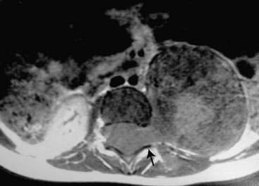
♦ Routine imaging studies include chest radiographs, ultrasonography, CT, and magnetic resonance imaging (MRI).
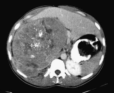
♦ The preferred diagnostic tool for initial imaging, staging, and the detection of metastases is CT (Fig. 34-1).