Chapter 68 Nail disorders
2. Why are nails important in medicine?
Nails are important because they are easily observable and serve as a window into the body. They are commonly affected by numerous internal (heart, liver, kidney) and external factors, including infectious agents, trauma, and drugs. Finally, nails may help in the differential diagnosis of closely related dermatologic disorders.
3. Do any systemic diseases have specific nail findings?
Many systemic diseases have characteristic, but not mutually exclusive, nail findings. Most nail changes are part of a symptom complex or a reaction pattern that may be extremely helpful in making a particular diagnosis (Table 68-1).
NAIL ABNORMALITY | AREA INVOLVED | ASSOCIATED DISEASE |
---|---|---|
Splinter hemorrhages | Bed | Bacterial endocarditis |
Mees’ lines | Plate | Arsenic exposure |
Muehrcke’s lines | Bed | Nephrotic syndrome |
Terry’s nails | Bed | Cirrhosis |
Half-and-half nails | Bed | Chronic renal failure |
Blue lunulae | Matrix | Wilson’s disease |
Red lunulae | Matrix | Rheumatoid arthritis |
Clubbing | Plate/matrix | Pulmonary disorders |
Spoon nails | Plate/matrix | Iron deficiency |
Nail fold telangiectasias | Nailfold | Scleroderma, systemic lupus |
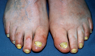
Yellow nails | Plate | Pulmonary disorders, sinusitis |
Scher RK, Daniel CR: Nails: therapy, diagnosis, surgery, ed 3: Philadelphia, 2005, WB Saunders.
4. What are Beau’s lines? How are they formed?
Beau’s lines represent the most common but least specific nail changes seen with systemic diseases. They are a forward-pointing, wedge-shaped depression in the nail plate of variable depth and obliquity. They occur when there is temporary cessation of nail growth or decreased deposition of nail plate by the nail matrix. Frequently, Beau’s lines are caused by mechanical trauma or diseases of the proximal nail fold. If all of the nails are affected at the same level, a systemic cause is indicated, while localized events (trauma) produce isolated lines.
5. What is a splinter hemorrhage?
A splinter hemorrhage results from the extravasation of blood from the longitudinally oriented vessels of the nail bed. The blood usually attaches to the overlying nail plate and moves distally with it. The occurrence of a hemorrhage close to the lunula and, simultaneously, in multiple nails correlates more directly with systemic disease.
6. Are splinter hemorrhages always associated with subacute bacterial endocarditis?
A commonly held, almost “sacred teaching” in medical school but rarely true. There is a myriad of causes, with subacute bacterial endocarditis representing only a small fraction. By far, simple trauma is the most common cause. Other known entities include drug reactions, general illness, vasculitis, and trichinosis, to name a few.
7. What is the difference between Mees’ lines and Muehrcke’s lines?
Mees’ lines represent single or multiple transverse white lines that occur in the nail plate and move distally as the nail grows out. They are classically thought to be caused by arsenic intoxication, but many severe systemic insults may initiate them. Muehrcke’s lines were described in 1956 by Robert C. Muehrcke in an article entitled “The Finger-nails in Chronic Hypoalbuminaemia.” These represent transverse, double, white lines that are an abnormality of the vascular bed, probably a localized edematous state secondary to the hypoalbuminemia. The underlying causes of these lines include the nephrotic syndrome, liver disease, and malnutrition.
Muehrcke RC: The finger-nails in chronic hypoalbuminaemia, Br Med J 9:1327–1328, 1956.
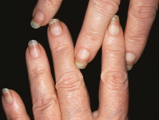
8. What are “half-and-half” nails, and with what internal disease are they associated?
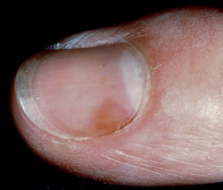
Half-and-half nails are characterized by apparent leukonychia (white color that disappears with pressure) that affects the proximal half of the nail (Fig. 68-1). Although it may be seen in normal individuals, it is associated with patients with chronic renal disease in approximately 10% of patients.
9. What is nail fold capillaroscopy? How is it useful?
It is the in vivo examination of nail fold and cuticle finger capillaries, with magnification, to detect variations in capillary patterns. Autoimmune connective tissue disorders can have subtle but distinct changes within these capillaries and the proximal nail fold. It can be useful in predicting which patients with Raynaud’s syndrome will likely develop scleroderma. In those with scleroderma, the severity of capillary lesions may correlate with the degree of multisystem organ disease. Patients with dermatomyositis have capillary changes along with thickened, ragged cuticles. In systemic lupus, paronychial inflammation may be prominent with dilated tortuous capillaries distinctive.
Figure 68-1. Half-and-half nails in a patient with chronic renal disease.
(Courtesy of the Fitzsimons Army Medical Center teaching files.)
Tosti A: The nail apparatus in collagen disorders, Semin Dermatol 10:71–76, 1991.
10. What is clubbing?
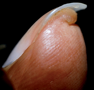
Clubbing refers to the increased bilateral curvature of the nails with proliferation of the soft tissues restricted to the distal phalanges (Fig. 68-2). It causes an increase in the emergence angle of the nail to equal or greater than 180 degrees. There are diverse causes of clubbing, including congenital or genetic factors, but 80% of clubbing is associated with respiratory ailments.
11. How is hypertrophic osteoarthropathy related to clubbing?
Clubbing may occur in association with hypertrophic osteoarthropathy, an uncommon but important entity. It consists of simple clubbing (including the toes), hypertrophy of the upper and lower extremities, peripheral neurovascular disease, acute burning bone pain, joint problems, and muscle weakness. More importantly, when complete, it is associated 90% of the time with malignant tumors of the chest.
Buy Membership for Dermatology Category to continue reading. Learn more here