192 Nail changes
Salient features
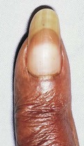
• Terry’s nail (Fig. 192.1): brownish-red distal transverse band; occurs normally in the elderly, also seen in cirrhosis and congestive cardiac failure
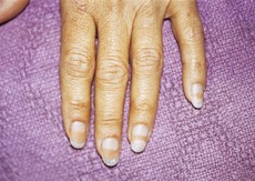
• Lindsay’s half-and-half nail (Fig. 192.2): distal brown band occupying about 25–50% of the nail bed, and seen in chronic renal failure
• Muehrcke’s nail: pale transverse bands resulting from oedema of the nail bed; seen in hypoalbuminaemia
• Blue lunula: in Wilson’s disease the normally white lunula becomes blue (p. 437); the lunula has the largest area in the fingers closest to the thumb; in the elderly it becomes smaller or absent.
• Disorders of the nail plate:
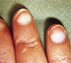
• Leukonychia (Fig. 192.3): whitish discoloration, which may be diffuse (liver disease, congenital, fungal), punctate or linear (Mees’ lines (Fig. 192.4) seen in arsenical poisoning) or vasculitic
• Onychomycosis: dystrophy, destruction of the nail caused by fungal or yeast infections (see Case 191)
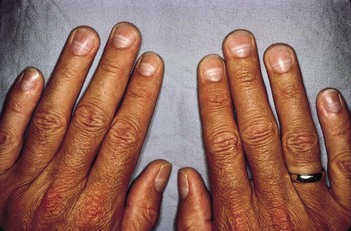
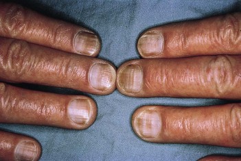
• Beau’s lines (Fig. 192.5): transverse depression occuring in severe systemic illness; it allows estimation of the date of the illness (normal nail grows at the rate of 0.1 mm/day and it takes approximately 3–4 months for the nail plate to grow out completely).