Myotomes
What is a myotome?
A myotome is defined as the group of muscles supplied by one spinal nerve root level. There are 31 pairs of spinal nerves (S2.13) each contributing to the innervations of many muscles (e.g. C5 innervates parts of supraspinatus, infraspinatus, deltoid and biceps). The muscles supplied by a single nerve root level are generally involved in a common muscle action/s and it is this muscle action that is assessed. The list below identifies a simplified version of the actions associated with each spinal nerve root level (Grieve 2004).
C1/C2 – neck flexion/neck extension (see Fig. 31.1)
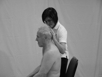
Figure 31.1 Testing for myotome C3.
C5 – shoulder abduction (see Fig. 31.2)
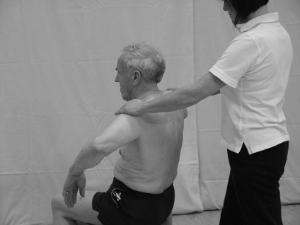
Figure 31.2 Testing for myotome C5.
C6 – elbow flexion or forearm supination
C7 – elbow extension or wrist flexion
C8 – thumb extension or adduction and ulna deviation
T1 – finger abduction/adduction
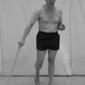
L3 – knee extension (see Fig. 31.3)
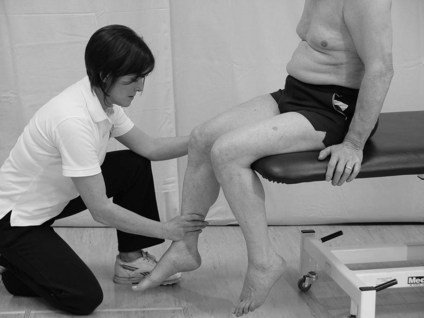
Figure 31.3 Testing for myotome L3.
L4 – dorsi flexion at the ankle
Why do I need to assess myotomes?
Assessing a myotome gives information related to nerve integrity, in other words, whether the nerve pathway from the spinal cord to the muscle is intact. The assessment evaluates the strength of the muscle contraction, however it should be remembered that a weak muscle could be the result of a lesion anywhere along the nerve pathway but also of the muscle itself. Knowledge of the muscles supplied by the spinal nerve root (myotome) and the peripheral nerve allows the therapist to differentiate between lesions of each (Petty 2006). A complete lesion of the peripheral nerve will lead to complete paralysis of the muscles innervated by that nerve. Therefore, weakness will be evident immediately on testing and muscle atrophy will occur over time. However, the presentation of a lesion to a single nerve root will be more difficult to recognize because the muscle itself will still be innervated by other unaffected root levels within the peripheral nerve. For example, the biceps muscle is supplied by the musculocutaneous nerve (C5/6/7). Therefore, any lesion affecting C6 nerve root will present as minor weakness because sufficient motor units can be recruited via the remaining root levels C5/7. However, if the therapist provides resistance over a period of time (5–10 seconds) the weakness may become evident.
 Caution
How do I assess a myotome?
Therapist
1. Identify a muscle action from the list above.
2. Resistance is applied by the therapist so that the muscles being assessed contract isometrically. Therefore no movement should occur at the joint which may confound the findings.
3. Where possible the test should be performed in middle range of the action (Figs 31.1–31.3).
4. The resistance should be brought on slowly to allow the patient to build up to the level of resistance offered by the therapist.
5. The amount of force applied should be appropriate to the size of the muscle being tested, especially for neck movements.
6. The isometric contraction should be held for approximately 5–10 seconds.
7. Where possible, the left and right sides should be compared simultaneously (Fig. 31.2).
Is there weakness evident in the action? Yes, there may be a deficit within the peripheral nervous system. If other spinal root levels appear intact the deficit may be localized to the root level being tested and should be confirmed using evidence from dermatome and reflex testing for the same level. However, if the distribution of loss is more generalized, this is unlikely to be the case.
Is the strength of contraction the same on both sides? No, this could indicate a unilateral deficit.