Myopic Choroidal Neovascular Membrane
Clinical Features:
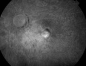
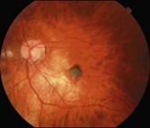
This type of CNV is usually well-circumscribed, heavily pigmented, and in a background of typical myopic changes (Fig. 9.2.1). It typically involved the fovea and is a type 2, or classic, CNV subtype. Its behavior tends to be less aggressive than CNV associated with wet age-related macular degeneration.
OCT Features:
Acutely, the CNV complex appears as a well-circumscribed area of mixed reflectivity in the subretinal space with overlying sub- and intraretinal fluid (Fig. 9.2.2, inset lower right). Sometimes, the presence of active CNV in high myopia can be difficult to discern, even with OCT. In this setting, activity may be recognized by subtle changes on serial exams (Figs 9.2.3 to 9.2.6). For such comparisons to be accurate, the scans that are taken at different times should be registered to assure the exact same region is being imaged over time. In the setting of myopic CNV, thickness maps are often fraught with segmentation artifact and cannot always be relied on to make treatment decisions.
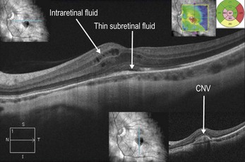
Figure 9.2.2 A horizontal line scan OCT (corresponding to Figure 9.2.1) of the fovea shows thin subretinal fluid with overlying intraretinal fluid at the edge of the lesion. The corresponding thickness map (inset, upper right) helps identify the affected area of thickened retina. A vertical line scan OCT (inset, bottom right) shows an elevated subretinal lesion with mixed reflectivity corresponding to the CNV.
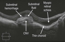
Figure 9.2.3 OCT shows a subretinal dome-shaped hyper-reflective area, which represents a myopic CNV. The distinction of the CNV from the overlying retina is blurred due to subretinal hemorrhage and subretinal fluid. These findings are more subtle in a myopic CNV in comparison to other forms of CNV. Characteristic myopic findings including a thin choroid and retinal schisis are also seen.
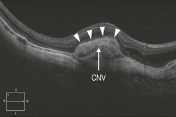
Figure 9.2.4 OCT (corresponding to Figure 9.2.3) shows a much more distinct border of the retina (arrowheads) and underlying myopic CNV one month following treatment with anti-VEGF therapy.
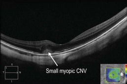
Figure 9.2.5 OCT of a small myopic CNV shows an ill-defined medium reflectivity subretinal elevation that obscures the photoreceptor and ELM layers and has poorly defined edges. (Courtesy of Caroline Baumal, MD.)
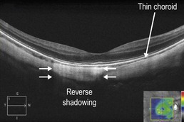
Figure 9.2.6 OCT (corresponding to Figure 9.2.5) shows complete resolution of the myopic CNV one month following treatment with intravitreal bevacizumab. (Courtesy of Caroline Baumal, MD.)